Stáhnout prezentaci
Prezentace se nahrává, počkejte prosím

1
HEMOKOAGULACE A FIBRINOLÝZY
VYŠETŘOVÁCÍ METODY U HEMOKOAGULACE A FIBRINOLÝZY Pavel Maruna Ústav patologické fyziologie LF UK

2
I. Fyziologie

3
Hemostáza = je mechanismus umožňující udržení oběhu po poruše cévní stěny. Sestává z těchto fází: 1. Klidová fáze – Udržování krve v tekutém stavu v cirkulaci 2. Aktivační fáze - Zástava krvácení v místě poranění pomocí hemostatické zátky 3. Fáze restituce- rekanalizace - Zajišťuje odstranění hemostatické zátky na konci reparačních procesů

4
Patho-physiological role
Hemostasis Hemostasis is an integral part of stress reaction inflammatory response Patho-physiological role X Protective role non-specific defense mechanism thrombosis / embolism atherosclerosis

5
Hemostasis Hemostasis as a physiological process must be: 1. Rapid
2. Localized 3. Reversible Inappropriate hemostasis: - Thrombosis / embolism - DIC (disseminated intra-vascular coagulation) - bleeding / blood loss
 - bleeding / blood loss.")
6
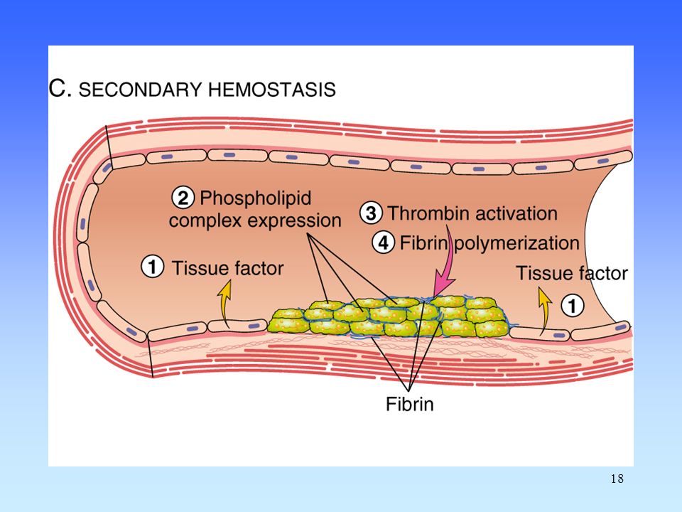
Hemostasis Vessel wall Endothelium Platelets Plasma coagulation system

7
Obecné mechanismy, popis regulace

8
Negativní (záporná) zpětná vazba
y…regulovaná veličina, v/v w…požadovaná hodnota e…regulační odchylka/ signál u…akční veličina d,n…poruchové veličiny U záporné zpětné vazby regulační odchylka e použitá k regulaci vznikne odečtením akční veličiny (-y) od požadované hodnoty (+w), e = w - y.
 od požadované hodnoty (+w), e = w - y.")
9
Pozitivní (kladná) zpětná vazba
y…regulovaná veličina, v/v w…požadovaná hodnota e…regulační odchylka/ signál u…akční veličina d,n…poruchové veličiny U kladné zpětné vazby regulační signál e vznikne přičtením akční veličiny (+y) k požadované hodnotě (+w), e = w + y.
 k požadované hodnotě (+w), e = w + y.")
10
Antithrombotic Properties
Endothelium Antithrombotic Properties Anti-platelet activities: Endothelium covers highly thrombogenic basal membrane Uninjured endothelium does not bind platelets PGI2 (prostaglandin) and NO (nitric oxide) from endothelium inhibit platelet binding ADPase counters the platelet aggregating effects of ADP
 and NO (nitric oxide) from endothelium inhibit platelet binding. ADPase counters the platelet aggregating effects of ADP.")
11
Antithrombotic Properties
Endothelium Antithrombotic Properties Anticoagulant activities: Heparin-like molecules ... activate anti-thrombin III (inactivates active proteases) Thrombomodulin ... changes specificity of thrombin (activates protein C , which inactivates factors Va and VIIIa tPA (tissue plasminogen activator) ... activates fibrinolysis via plasminogen to plasmin
 Thrombomodulin ... changes specificity of thrombin (activates protein C , which inactivates factors Va and VIIIa. tPA (tissue plasminogen activator) ... activates fibrinolysis via plasminogen to plasmin.")
12
Prothrombotic Properties
Endothelium Prothrombotic Properties Synthesis of von Willebrand factor Release of tissue factor Production of PAI (plasminogen activator inhibitors) Membrane phospholipids bind and facilitate activation of clotting factors via Ca2+ bridges
 Membrane phospholipids bind and facilitate activation of clotting factors via Ca2+ bridges.")
13
Endothelium Antithrombogenic Thrombogenic Vessel injury
(Favors fluid blood) (Favors clotting)
 (Favors clotting)")
14
Vasoconstriction Primary hemostasis Secondary hemostasis Fibrinolysis

15
ECM (=ExtraCellular Matrix)
")
16
TXA2 (thromboxane A2, lipid)
")
17
Gp – G-protein coupled receptors

19
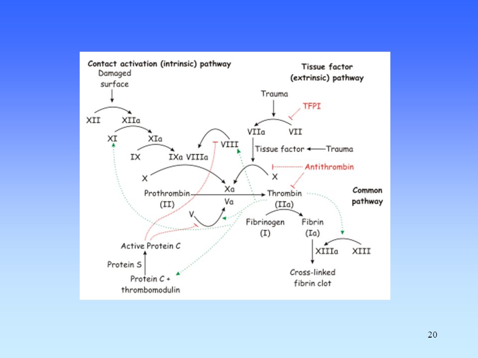
Coagulation Intrinsic pathway Extrinsic Pathway XIIa XIa TF
Prothrombin IXa VIIa VIII VIIIa Xa Va V Soft clot Thrombin Fibrinogen Fibrin XIIIa Hard clot Fibrin

21
Coagulation Enzymatic cascade (amplification) Several serine proteases
Produced by liver (most) Require vitamin K (several, 2, 7, 9, 10, C, S) Requires Ca 2+ (the same, 2, 7, 9, 10, C, S) 3 protein cofactors (not enzymes) Reversible (via production of plasmin)
 Require vitamin K (several, 2, 7, 9, 10, C, S) Requires Ca 2+ (the same, 2, 7, 9, 10, C, S) 3 protein cofactors (not enzymes) Reversible (via production of plasmin)")
22
Coagulation

23
Coagulation

24
Coagulation Thrombin Fibrinogen Fibrin

25
Coagulation Prothrombin Xa Va Thrombin Fibrinogen Fibrin

26
Coagulation Extrinsic Pathway TF Prothrombin VIIa Xa Va Thrombin
Fibrinogen Fibrin

27
Coagulation Intrinsic pathway Extrinsic Pathway XIIa XIa TF
Prothrombin IXa VIIa VIIIa Xa Va Thrombin Fibrinogen Fibrin

28
Coagulation Intrinsic pathway Extrinsic Pathway XIIa XIa TF
Prothrombin IXa VIIa VIIIa Xa Va Soft clot Thrombin Fibrinogen Fibrin XIIIa Hard clot Fibrin

29
Coagulation Intrinsic pathway Extrinsic Pathway XIIa XIa TF
Prothrombin IXa VIIa VIII VIIIa Xa Va V Soft clot Thrombin Fibrinogen Fibrin XIIIa Hard clot Fibrin

30
Revised tissue factor pathway
Extrinsic Pathway IX TF Prothrombin (II) IXa VIIa Xa Thrombin (IIa) New: Production of IXa Interaction of intrinsic and extrinsic pathways
 IXa. VIIa. Xa. Thrombin (IIa) New: Production of IXa. Interaction of intrinsic and extrinsic pathways.")
31
Revised tissue factor pathway
IX TF Prothrombin (II) IXa VIIa Xa TFPI TFI Thrombin (IIa) New: TFPI = Tissue Factor Pathway Inhibitor ... inhibition of Xa and VIIa
 IXa. VIIa. Xa. TFPI. TFI. Thrombin (IIa) New: TFPI = Tissue Factor Pathway Inhibitor. ... inhibition of Xa and VIIa.")
32
Revised tissue factor pathway
TFPI is protease inhibitor 34 and 41 kD forms in plasma (C-term truncation) Activities: - direct inhibition of Xa - inhibition VIIa-TF complex in a [Xa]-dependent manner - binding to LDL, HDL and Lp (a) ~10% present in platelets (endothelium also) IX TF Prothrombin (II) IXa VIIa Xa TFPI TFI New: TFPI = Tissue Factor Pathway Inhibitor ... inhibition of Xa and VIIa
 Activities: - direct inhibition of Xa. - inhibition VIIa-TF complex in a [Xa]-dependent manner. - binding to LDL, HDL and Lp (a) ~10% present in platelets (endothelium also) IX. TF. Prothrombin (II) IXa. VIIa. Xa. TFPI. TFI. New: TFPI = Tissue Factor Pathway Inhibitor. ... inhibition of Xa and VIIa.")
33
Revised tissue factor pathway
Net results: Production of IXa Production of small amounts of thrombin (IIa) No or only little fibrin formed!
 No or only little fibrin formed!")
34
Revised tissue factor pathway
VIIa forms via binding of VII to TF VIIa activates some XXa Xa converts a small amount of II to IIa; this thrombin is used to produce small amts of VIIIa and Va As the concentration of TF-VIIa-Xa-IIa increases, TFPI inactivates this complex stopping further production of thrombin. IXa, with VIIIa (produced as above), produces Xa; this Xa with Va produces new thrombin; this thrombin produces more VIIIa and Va and then we get lots of thrombin and fibrin.
, produces Xa; this Xa with Va produces new thrombin; this thrombin produces more VIIIa and Va and then we get lots of thrombin and fibrin.")
35
Revised tissue factor pathway
IX TF Prothrombin IXa VIIa VIII VIIIa Xa Va V Soft clot Thrombin Fibrinogen Fibrin XIIIa Hard clot Fibrin

36
Revised tissue factor pathway
VIII Thrombin (IIa) Va VIIIa
 Va. VIIIa.")
37
Role of vitamin K Factors II, VII, IX, X, proteins C and S
require a post-translational modification (PTM) before their activation This PTM requires vitamin K This PTM involves the addition of a COO- to certain Glu residues in the clotting factors resulting in the formation of several gamma-carboxy glutamates
 before their activation. This PTM requires vitamin K. This PTM involves the addition of a COO- to. certain Glu residues in the clotting factors. resulting in the formation of several. gamma-carboxy glutamates.")
38
Role of vitamin K

39
Physiologic inhibitors of coagulation
Antithrombin III SERPIN Activated Protein C + protein S Inactivates Va and VIIIa (via proteolysis) mutation: Factor V Leiden (APC resistance) Thrombomodulin Binds to thrombin Decreases ability to produce fibrin Increases ability to activate Protein C
 mutation: Factor V Leiden (APC resistance) Thrombomodulin. Binds to thrombin. Decreases ability to produce fibrin. Increases ability to activate Protein C.")
40
Non-physiologic inhibitors of coagulation
Vitamin K antagonists (in vivo only) Ca chelators (in vitro only) EDTA Citrate Oxalate Heparin (in vivo and in vitro)
 Ca chelators. (in vitro only) EDTA. Citrate. Oxalate. Heparin. (in vivo and in vitro)")
41
Fibrinolysis ... Clot removal

42
Fibrinolysis Plasmin Fibrin Fibrin Split Products (FSP)
")
43
Fibrinolysis Plasminogen bacterial enzymes (streptokinase) tPA uPA
Fibrin Split Products (FSP)
")
44
Inhibitors of fibrinolysis
Plasminogen PAI tPA a2-antiplasmin ... SERPINs Plasmin Fibrin Fibrin Split Products (FSP)
")
45
Fibrinolysis

46
II. Pathology

47
Coagulopathies Congenital Acquired

48
Coagulopathies Congenital Acquired Hemophilia A ... f VIII
Hemophilia B ... f IX Hemophilia C ... f XI Dys- / A- fibrinogenemia F V defic. (parahemophilia) F XIII defic. APC resistance
 F XIII defic. APC resistance.")
49
Coagulopathies Congenital Acquired Liver proteosynthesis
Vitamin K defic. - obstructive icterus - intestin. resorption Anticoagulant therapy - Dicumarol - Heparin

50
Vasculopathies Congenital Acquired Mb. Rendu-Osler-Weber
= hereditary hemorrhagic teleangiectasia AD, TGFbeta1 rec. Ehlers-Danlos Sy. = defects in collagen synthesis Purpura Henoch-Schönlein Scorbut Steroid purpura Purpura simplex and senilis

51
Rizikové faktory a příklady žilních uzávěrů
Rizikové faktory vzniku žilní trombózy: -útlak cévy zvnějšku (např. trombóza hlubokých žil levé dolní končetiny je cca 3x častější než pravé dolní končetiny…. Proč?) -hyperviskozita (dehydratace, polyglobulie, leukémie, hyperfibrinogenémie aj.) -městnání krve v žilním systému (deficientní chlopně žil, útlak žil zvnějšku) -imobilita -obezita -nadměrná aktivace sekundární hemostázy (např. u infekcí, zánětů, malignit, v těhotenství) -samostatnou kapitolou jsou vrozené trombofilní stavy. Příklady žilních uzávěrů: -flebotrombóza DK= trombóza hlubokých žil dolních končetin -tromboflebitida DK= trombóza povrchových žil dolních končetin -plicní trombembolismus -trombóza viscerálních žil (např. trombóza vrátnicové žíly, trombóza jaterních žil: tzv. Budd-Chiariho syndrom) -Trousseauův příznak (migrující tromboflebitida u nádorových onemocnění) -samostatnou kapitolou jsou žilní uzávěry u chronických hemolytických anémií a klonálních poruch krvetvorby (MPN, PNH)
 -hyperviskozita (dehydratace, polyglobulie, leukémie, hyperfibrinogenémie aj.) -městnání krve v žilním systému (deficientní chlopně žil, útlak žil zvnějšku) -imobilita -obezita -nadměrná aktivace sekundární hemostázy (např. u infekcí, zánětů, malignit, v těhotenství) -samostatnou kapitolou jsou vrozené trombofilní stavy. Příklady žilních uzávěrů: -flebotrombóza DK= trombóza hlubokých žil dolních končetin -tromboflebitida DK= trombóza povrchových žil dolních končetin -plicní trombembolismus -trombóza viscerálních žil (např. trombóza vrátnicové žíly, trombóza jaterních žil: tzv. Budd-Chiariho syndrom) -Trousseauův příznak (migrující tromboflebitida u nádorových onemocnění) -samostatnou kapitolou jsou žilní uzávěry u chronických hemolytických anémií a klonálních poruch krvetvorby (MPN, PNH)")
52
Trombóza hlubokých žil levé dolní končetiny vzniká cca 3x častěji než trombóza pravé dolní končetiny

53
Genetic examination Hemophilia A X-linked recessive 1 :

54
Clinical signs Hemophilia Large hemorrhage after a small injury
Arthral hemorrhage Secondary arthropathy Hemophilia

55
Clinical signs Thrombocytopenia Petechiae, pigmentation

56
Clinical signs Henoch-Schonlein

57
Clinical signs F XIII deficiency Late bleeding Keloid scarring

58
Deep venous thrombosis
Clinical signs Deep venous thrombosis Pulmonary embolism

59
Diagnostics and monitoring
III. Diagnostics and monitoring

60
Standard tests in Faculty General Hospital
Quick time, INR 0,8 - 1,2 Act.Part.Thromb.Time s Thrombin time s Fibrinogen g/l Antithrombin III > 70% Ethanol test neg. D-dimers (FDP) neg.
 neg.")
61
Prothrombin Time (Quick test)
Principle: Stimulation of extrinsic (main) coag. system Citrate plasma ... add TF (in excesive amount) + CaCl2 ... fibrin fibre Normal: PT = s INR = (PTP)ISI / PTN ISI = international index of sensitivity of used thromboplastin (commonly > 1) Prolongation: defic. vit. K dep. FII, VII, X, Fbg Usage: screening, monitoring of oral anticoagulants, liver proteosynthesis Normal range INR 0,8 - 1,2 Therapeutic range INR = 2,5 - 4,5 Surgery INR < 1,6
 coag. system. Citrate plasma ... add TF (in excesive amount) + CaCl2 ... fibrin fibre. Normal: PT = s. INR = (PTP)ISI / PTN. ISI = international index of sensitivity of used thromboplastin (commonly > 1) Prolongation: defic. vit. K dep. FII, VII, X, Fbg. Usage: screening, monitoring of oral anticoagulants, liver proteosynthesis. Normal range INR 0,8 - 1,2. Therapeutic range INR = 2,5 - 4,5. Surgery INR < 1,6.")
62
APTT, Activated partial thromboplastin time
Principle: Stimulation of intrinsic (contact) way of coag. system Citrate plasma ... add contact activator (e. g. kaolin) + CaCl2 ... fibrin fibre
 way of coag. system. Citrate plasma ... add contact activator (e. g. kaolin) + CaCl2 ... fibrin fibre.")
63
APTT, Activated partial thromboplastin time
Principle: Stimulation of intrinsic (contact) way of coag. system Citrate plasma ... add contact activator (e. g. kaolin) + CaCl2 ... fibrin fibre Normal: APTT = s Prolongation: defic. of VII, V, X, XII, VIII, XI, IX (hemophilia A,B,C), Fbg, FDP Shortening: prothrombotic status Usage: screening, diagnostics of coagul. deficits, monitoring of heparin therapy Therapeutic range 1,2 - 2,5 x
 way of coag. system. Citrate plasma ... add contact activator (e. g. kaolin) + CaCl2 ... fibrin fibre. Normal: APTT = s. Prolongation: defic. of VII, V, X, XII, VIII, XI, IX (hemophilia A,B,C), Fbg, FDP. Shortening: prothrombotic status. Usage: screening, diagnostics of coagul. deficits, monitoring of heparin therapy. Therapeutic range 1,2 - 2,5 x.")
64
Lee-White test Cloting time of whole blood
Whole blood without anticoagulants (CaCl2) ... polystyrene or glass tube, 37°C ... spontaneous stimulation of intrinsic Normal: min. Usage: Basic, rough orientation in acute status
 ... polystyrene or glass tube, 37°C ... spontaneous stimulation of intrinsic. Normal: min. Usage: Basic, rough orientation in acute status.")
65
Thrombin Time Whole blood without anticoagulants (CaCl2) ... add thrombin in standard amount, 37°C ... fibrin fibre Normal: s Prolongation: Fbg (acute stage of DIC) antithrombins fibrinolysis Usage: DIC monitoring of fibrinolytic therapy
 antithrombins. fibrinolysis. Usage: DIC. monitoring of fibrinolytic therapy.")
66
Fibrinogen, Fbg Normal plasma levels = 2 - 4 g /l
Functional of immunological detection High: Inflammation DM Smoking Low: Low synthesis (congenital or low liver function) Consumption (DIC) Hypofibrinogenemia Dysfibrinogenemia
 Consumption (DIC) Hypofibrinogenemia. Dysfibrinogenemia.")
67
FDP Total degradation products of fibrin(-ogen)
ELISA or aglutination semiquantitative methods High: Recent coagulation activity (thrombo/ embolism, bleeding, surgery, DIC ...) High sensitivity, low specificity
 High sensitivity, low specificity.")
68
Paracoagulation tests (Ethanol, Protamin)
Principle: Ethanol catalyzes conversion of fibrin monomers + PDP fibrin polymers Low sensitivity and specificity Usage: 1st stage of DIC

69
Duke test Duke, 1910 Estimation of bleeding time
Time of spontaneous cutoff of bleeding after standard puncture to auricle of ear Limits: min., or min. (depends on methods) Prolongation - Disturbance of primary hemostasis: Plt < or Plt dysfunction, vW disease
 Prolongation - Disturbance of primary hemostasis: Plt < or Plt dysfunction, vW disease.")
70
Rumpel - Leede test Capillary resistance
Number of petechia on forearm (area 4 x 4 cm) after a standard pressure (ruff 10,5 kPa for 10 min.) or after underpressure (Brown, 1949) Limits: > 5 petechia ... higher capillary fragility (e.g. hereditary purpura Weber-Rendu-Osler)
 after a standard pressure (ruff 10,5 kPa for 10 min.) or after underpressure (Brown, 1949) Limits: > 5 petechia ... higher capillary fragility. (e.g. hereditary purpura Weber-Rendu-Osler)")
71
Presumable results Diagnosis Plt Duke APTT Quick TT
Thrombocytopenia N N N Hemophilia A N N N N Hemophilia B N N N N Hemophilia C N N N N vWd N N / N N

72
Presumable results Diagnosis Plt Duke APTT Quick TT
F V defic. N N N F II defic. N N N N F VII defic. N N N N Warfarin / vit. K def. N N N Heparin i. v. N N / N / Heparin s. c. N N N N N

73
Presumable results Diagnosis Plt Ethan APTT Quick TT
DIC 1st stage + N DIC 2nd stage -

74
Standard tests in Faculty General Hospital
Quick time, INR 0,8 - 1,2 APTT s Thrombin time s Fibrinogen g/l Antithrombin III > 70% Ethanol test neg. D-dimers (FDP) neg.
 neg.")
75
Risc factors and examples of VTE (venous thrombo-embolism)
Risc factors: -vessel oppression (e.g. phlebo-thrombosis of left lower extremity is circa 3 times more common than phlebo-thrombosis of right lower extremity ….Why is that so?) -dehydration -hyperviscosity -stasis syndrom (e.g. right heart insufficiency, long airplane flight) -immobility -obesity -activation of secondary hemostasis, e.g. Inflammation, infection, trauma, malignancies -inborn hypercoagulable states Examples: -phlebothrombosis of deep veins of lower extremities -thrombophlebitis of superficial veins of lower extremities -lung thrombembolism -thrombosis of large visceral veins (e.g. thrombosis of vena portae, hepatic vein thrombosis= Budd-Chiari syndrome) -Trousseau symptom (migratory thrombophlebitis in malignancies) -thrombotic complications in chronic hemolytic anemias (sickle cell anemia, thalassemias) and clonal disorders of hematopoiesis (MPN, PNH)
 -dehydration -hyperviscosity -stasis syndrom (e.g. right heart insufficiency, long airplane flight) -immobility -obesity -activation of secondary hemostasis, e.g. Inflammation, infection, trauma, malignancies -inborn hypercoagulable states Examples: -phlebothrombosis of deep veins of lower extremities -thrombophlebitis of superficial veins of lower extremities -lung thrombembolism -thrombosis of large visceral veins (e.g. thrombosis of vena portae, hepatic vein thrombosis= Budd-Chiari syndrome) -Trousseau symptom (migratory thrombophlebitis in malignancies) -thrombotic complications in chronic hemolytic anemias (sickle cell anemia, thalassemias) and clonal disorders of hematopoiesis (MPN, PNH)")
Podobné prezentace