Stáhnout prezentaci
Prezentace se nahrává, počkejte prosím

1
M. Šmajerová, T. Andrašina, Š. Bohatá Ježíši důvěřuji v Tebe

2
dřívější detekce nových primárních a metastatických malignit, charakterizace ložiska, histologický grading, rychlejší a komplexnější hodnocení léčebné odpovědi, dřívější záchyt relapsu Využití k hodnocení: GIST, HCC, meta CRC, Ca prsu, Ca ledvin Lim KS. Diffusion-weighted MRI of hepatocellular carcinoma in cirrhosis. Clinical Radiology. 2014;69(1):1-10
:1-10.")
3
Chemoterapie → cytostatický účinek → → destrukce buněk → zmenšení velikosti Biologická léčba → antiangiogenní účinek → → snížení perfuze → velikost stejná http://www.avastin.com/patient/about/rgbm-difference-from-chemotherapy Biological therapy

4
Růst nových cév Potřebná u tumorů větších než 1-2mm Stimulem je hypoxie, v nádoru narušená regulace Výsledek - chaotická cévní struktura 1. Bowden DJ, Barrett T. Angiogenesis Imaging in Neoplasia. Journal of Clinical Imaging Science. 2011;1(1):1-7 2 Miller JC, Pien HH, Sahani D, Sorensen AG, Thrall JH. Imaging Angiogenesis: Applications and Potential for Drug Development. JNCI: Journal of the National Cancer Institute. 2005;97(3):172-187
:1-7 2 Miller JC, Pien HH, Sahani D, Sorensen AG, Thrall JH. Imaging Angiogenesis: Applications and Potential for Drug Development. JNCI: Journal of the National Cancer Institute. 2005;97(3):")
5
Histologická metoda „Zlatý standard“ v kvantifikování angiogeneze 1. Bowden DJ, Barrett T. Angiogenesis Imaging in Neoplasia. Journal of Clinical Imaging Science. 2011;1(1):1-7 2. Nico B, Benagiano V, Mangieri D, Maruotti N, Vacca A, Ribatti D. Evaluation of microvascular density in tumors: pro and contra. Histology and Histopathology. 2008;23(5):601-607 Nevýhody: biopsie – invasivní výkon nelze hodnotit funkční parametry malý vzorek
: Nico B, Benagiano V, Mangieri D, Maruotti N, Vacca A, Ribatti D. Evaluation of microvascular density in tumors: pro and contra. Histology and Histopathology. 2008;23(5): Nevýhody: biopsie – invasivní výkon nelze hodnotit funkční parametry malý vzorek.")
6
MR: DCE-MR, DWI-MR, MR elastografie, MR spektroskopie CT: Perfuzní CT, Dual-energy CT UZ: Doppler, CEUS NM: PET, SPECT -používané v klinické praxi

7
Difuze – náhodný pohyb molekul vody v prostředí DWI měří difuzi – volnost pohybu molekul vody v tkáni Detekce, charakterizace, histologický grading, hodnocení léčebné odpovědi Lim KS. Diffusion-weighted MRI of hepatocellular carcinoma in cirrhosis. Clinical Radiology. 2014;69(1):1-10.
:")
8
1. Sahani DV, Jiang T, Hayano K, et al. Magnetic resonance imaging biomarkers in hepatocellular carcinoma: association with response and circulating biomarkers after sunitinib therapy. Journal of Hematology & Oncology. 2013;6(1):51. 2. Cui Y, Zhang X-P, Sun Y-S, Tang L, Shen L. Apparent Diffusion Coefficient: Potential Imaging Biomarker for Prediction and Early Detection of Response to Chemotherapy in Hepatic Metastases. Radiology. 2008;248(3):894-900 Sahani, 2013 „There was an increase in median tumor ADC value after 2-week postsunitinib and in median tumor thrombus ADC value.“ Cui, 2008 „An early increase in ADCs (day 3 or 7) was observed in responding lesions but not in nonresponding lesions (P.002).“
: Cui Y, Zhang X-P, Sun Y-S, Tang L, Shen L. Apparent Diffusion Coefficient: Potential Imaging Biomarker for Prediction and Early Detection of Response to Chemotherapy in Hepatic Metastases. Radiology. 2008;248(3): Sahani, 2013 „There was an increase in median tumor ADC value after 2-week postsunitinib and in median tumor thrombus ADC value. Cui, 2008 „An early increase in ADCs (day 3 or 7) was observed in responding lesions but not in nonresponding lesions (P.002). .")
9
Modifikace T2 sekvencí pomocí rozfázování a opětovného zfázování spinů ◦ Vysoká difuze → spiny se volně pohybují → → nedokonalé zfázování → nízký signál (facilitace) ◦ Nízká difuze → spiny se nepohybují → → dokonalé zfázování → vysoký signál (restrikce) 1.Padhani A. Diffusion Magnetic Resonance Imaging in Cancer Patient Management. Seminars in Radiation Oncology. 2011;21(2):119-140 2.2. ECR 2012 / C-1677 / Diffusion-weighted whole-body imaging with background body signal suppression (DWIBS): a useful tool in daily practice - EPOS TM
: ECR 2012 / C-1677 / Diffusion-weighted whole-body imaging with background body signal suppression (DWIBS): a useful tool in daily practice - EPOS TM.")
10
Přídatný magnetický gradient - „B hodnoty“ [s/mm 2 ] Aparentní difusní koeficient (ADC) [mm 2 /s ] → ADC mapy ◦ minimálně 2 měření s různou B hodnotou 1. Padhani A. Diffusion Magnetic Resonance Imaging in Cancer Patient Management. Seminars in Radiation Oncology. 2011;21(2):119-140 2. Heijmen L. Diffusion-weighted MR imaging in liver metastases of colorectal cancer: reproducibility and biological validation. European Radiology. 2013; 23:748–756
![ Přídatný magnetický gradient - „B hodnoty [s/mm 2 ] Aparentní difusní koeficient (ADC) [mm 2 /s ] → ADC mapy ◦ minimálně 2 měření s různou B hodnotou 1.](http://images.slideplayer.cz/35/10393428/slides/slide_10.jpg "Padhani A. Diffusion Magnetic Resonance Imaging in Cancer Patient Management. Seminars in Radiation Oncology. 2011;21(2): Heijmen L. Diffusion-weighted MR imaging in liver metastases of colorectal cancer: reproducibility and biological validation. European Radiology. 2013; 23:748–756.")
11
Padhani A. Diffusion Magnetic Resonance Imaging in Cancer Patient Management. Seminars in Radiation Oncology. 2011;21(2):119-140
:")
12
T1 po k.l.

13
PŘED PO PředPo Rozdíl ADC [μm 2 x s]1,131,54 +36% ADCb600 ADC
![PŘED PO PředPo Rozdíl ADC [μm 2 x s]1,131,54 +36% ADCb600 ADC](http://images.slideplayer.cz/35/10393428/slides/slide_13.jpg "PŘED PO PředPo Rozdíl ADC [μm 2 x s]1,131,54 +36% ADCb600 ADC")
14
Bez potřeby k.l. Relativně jednoduché skenovací protokoly Širší dostupnost Kvantitativní analýza Dobrá reproducibilita Bowden DJ, Barrett T. Angiogenesis Imaging in Neoplasia. Journal of Clinical Imaging Science. 2011;1(1):1-7
:1-7.")
15
Vyšší cena Artefakty Zobrazování malých lézí (<1cm) a ložisek těsně pod bránicí Chybí definovaná kritéria v hodnocení léčebné odpovědi Bowden DJ, Barrett T. Angiogenesis Imaging in Neoplasia. Journal of Clinical Imaging Science. 2011;1(1):1-7
:1-7.")
16
Metoda pro hodnocení mikrovaskularizace Umožňuje posoudit nejen celkovou perfuzi, ale také odlišit arteriální a portální složku Liver Imaging Research Program | BIRC, http://www.birc.ca/liver-imaging-research-program

17
1. Jiang T, Kambadakone A, et al. Monitoring response to antiangiogenic treatment and predicting outcomes in advanced hepatocellular carcinoma using image biomarkers, ct perfusion, tumor density, and tumor size (RECIST). Investigative Radiology. 2012;47(1):11-17 2. Sabir A, Schor-Bardach R, Wilcox CJ, et al. Perfusion MDCT Enables Early Detection of Therapeutic Response to Antiangiogenic Therapy. American Journal of Roentgenology. 2008;191(1):133-139. Sabir, 2008 „In the sorafenib group, the mean blood flow was significantly lower at day 4, day 9 and day 14 compared with day 0 (p< 0.05).“ Jiang, 2012 „On days 10 to 12 after initiation of bevacizumab, significant decrease in CTp parameters was noted (P < 0.005).“
. Investigative Radiology. 2012;47(1): Sabir A, Schor-Bardach R, Wilcox CJ, et al. Perfusion MDCT Enables Early Detection of Therapeutic Response to Antiangiogenic Therapy. American Journal of Roentgenology. 2008;191(1): Sabir, 2008 „In the sorafenib group, the mean blood flow was significantly lower at day 4, day 9 and day 14 compared with day 0 (p< 0.05). Jiang, 2012 „On days 10 to 12 after initiation of bevacizumab, significant decrease in CTp parameters was noted (P < 0.005). .")
18
Nativní CT Rozsah 4-20cm Bolus jodové k.l. i.v. (40-50ml rychlostí 5- 10ml/s) Akvizice: ◦ First pass study – prvních 45-60s po 1-2s ◦ Opožděné zobrazování – ≤3min po 5-15s Analýza dat, korekce pohybu ROI (ložisko, játra, aorta, v.portae, slezina) 1. Goh V, Gourtsoyianni S, Koh D-M. Functional Imaging of the Liver. Seminars in Ultrasound, CT and MRI. 2013;34(1):54-65.. 2. Miles K a. Perfusion CT for the assessment of tumour vascularity: Which protocol? British Journal of Radiology. 2003;76(SPEC.ISS.1):S36-S42.
 Akvizice: ◦ First pass study – prvních 45-60s po 1-2s ◦ Opožděné zobrazování – ≤3min po 5-15s Analýza dat, korekce pohybu ROI (ložisko, játra, aorta, v.portae, slezina) 1. Goh V, Gourtsoyianni S, Koh D-M. Functional Imaging of the Liver. Seminars in Ultrasound, CT and MRI. 2013;34(1): Miles K a. Perfusion CT for the assessment of tumour vascularity: Which protocol. British Journal of Radiology. 2003;76(SPEC.ISS.1):S36-S42..")
19
Kim SH, Kamaya A, Willmann JK. CT Perfusion of the Liver: Principles and Applications in Oncology. Radiology. 2014;272(2):322-344 Průtok krve - blood flow BF [ml/100ml/min] Objem krve – blood volume BV [ml/100ml] Střední tranzitní čas – mean transit time MTT [s] Čas do max. nasycení – time to peak TTP [s] Permeabilita – PMB [ml/100g/min] Arteriální a portální perfuze [ml/100ml/min] Jaterní perfusní index HPI [%]
: Průtok krve - blood flow BF [ml/100ml/min] Objem krve – blood volume BV [ml/100ml] Střední tranzitní čas – mean transit time MTT [s] Čas do max. nasycení – time to peak TTP [s] Permeabilita – PMB [ml/100g/min] Arteriální a portální perfuze [ml/100ml/min] Jaterní perfusní index HPI [%].")
20
Kim SH, Kamaya A, Willmann JK. CT Perfusion of the Liver: Principles and Applications in Oncology. Radiology. 2014;272(2):322-344 PŘED BF [ml/100ml/ min] 62,1 BV [ml/100ml] 14,9 PMB [ml/100g/ min] 26,4 HPI [%]99,8
: PŘED BF [ml/100ml/ min] 62,1 BV [ml/100ml] 14,9 PMB [ml/100g/ min] 26,4 HPI [%]99,8.")
21
Kim SH, Kamaya A, Willmann JK. CT Perfusion of the Liver: Principles and Applications in Oncology. Radiology. 2014;272(2):322-344 PO PŘEDPO Rozdíl [%] BF [ml/100ml/ min] 62,129,6 -52,3 BV [ml/100ml] 14,91,3 -90,9 PMB [ml/100g/ min] 26,420,2 -23,2 HPI [%]99,863,0 -36,9
: PO PŘEDPO Rozdíl [%] BF [ml/100ml/ min] 62,129,6 -52,3 BV [ml/100ml] 14,91,3 -90,9 PMB [ml/100g/ min] 26,420,2 -23,2 HPI [%]99,863,0 -36,9.")
22
Relativně jednoduchá technika Možnost kvantitativní analýzy Výborné prostorové rozlišení Dobrá reproducibilita Časné mikrovaskulární změny 1. Bowden DJ, Barrett T. Angiogenesis Imaging in Neoplasia. Journal of Clinical Imaging Science. 2011;1(1):1-7 2. Jiang T, Zhu AX, Sahani DV. Established and novel imaging biomarkers for assessing response to therapy in hepatocellular carcinoma. Journal Of Hepatology. 2013;58(1):169-177
: Jiang T, Zhu AX, Sahani DV. Established and novel imaging biomarkers for assessing response to therapy in hepatocellular carcinoma. Journal Of Hepatology. 2013;58(1):")
23
Vyšší radiační zátěž Alergické reakce a toxicita jodové k.l. Nejsou standardizované protokoly Chybí definovaná kritéria v hodnocení léčebné odpovědi 1. Bowden DJ, Barrett T. Angiogenesis Imaging in Neoplasia. Journal of Clinical Imaging Science. 2011;1(1):1-7 2. Jiang T, Zhu AX, Sahani DV. Established and novel imaging biomarkers for assessing response to therapy in hepatocellular carcinoma. Journal Of Hepatology. 2013;58(1):169-177
: Jiang T, Zhu AX, Sahani DV. Established and novel imaging biomarkers for assessing response to therapy in hepatocellular carcinoma. Journal Of Hepatology. 2013;58(1):")
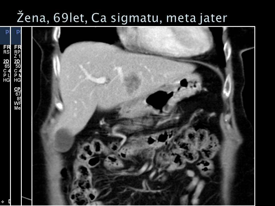
24
UZ vyšetření s aplikací speciální intravenózní k.l. Mikrobublinky plynu vel. 2-5μm stabilizované obalem z fosfolipidů nebo polymerů Bowden DJ, Barrett T. Angiogenesis Imaging in Neoplasia. Journal of Clinical Imaging Science. 2011;1(1):1-7
:1-7.")
25
1. Zocco M, Garcovich M, Lupascu A, et al. Early prediction of response to sorafenib in patients with advanced hepatocellular carcinoma: The role of dynamic contrast enhanced ultrasound. Journal of Hepatology. 2013;59(5):1014-1021 1 2. Sugimoto K, Moriyasu F, Saito K, et al. Hepatocellular carcinoma treated with sorafenib: early detection of treatment response and major adverse events by contrast-enhanced US. Liver International. 2013;33(4):605-615 Zocco, 2013 „The percentage variation at day 15 significantly correlated with response.“ Sugimoto, 2013 „Tumour perfusion parameters were statistically significant, the most relevant for tumour is response on day 7, the most relevant for PFS and OS.“
: Sugimoto K, Moriyasu F, Saito K, et al. Hepatocellular carcinoma treated with sorafenib: early detection of treatment response and major adverse events by contrast-enhanced US. Liver International. 2013;33(4): Zocco, 2013 „The percentage variation at day 15 significantly correlated with response. Sugimoto, 2013 „Tumour perfusion parameters were statistically significant, the most relevant for tumour is response on day 7, the most relevant for PFS and OS. .")
26
Lassau N, Chami L, Chebil M, et al. Advanced hepatocellular carcinoma: Early evaluation of response to bevacizumab therapy at dynamic contrast-enhanced us with quantification- preliminary results. Radiology. 2011;258(1):291-300 Lassau, 2011 „Dynamic US can be used to quantify dynamic changes in tumor vascularity as early as 3 days after bevacizumab administration in patients with HCC. These early changes in tumor perfusion may be predictive of tumor response at 2 months, progression-free survival, and overall survival“
: Lassau, 2011 „Dynamic US can be used to quantify dynamic changes in tumor vascularity as early as 3 days after bevacizumab administration in patients with HCC. These early changes in tumor perfusion may be predictive of tumor response at 2 months, progression-free survival, and overall survival .")
27
Nativní vyšetření, detekce ložiska Správné nastavení přístroje, harmonické zobrazení Bolus i.v. k.l. (2,4ml SonoVue®) Kvalitativní hodnocení: Leen E. The role of contrast-enhanced ultrasound in the characterisation of focal liver lesions. European Radiology. 2001;11(SUPPL.3):E27-E34.
 Kvalitativní hodnocení: Leen E. The role of contrast-enhanced ultrasound in the characterisation of focal liver lesions. European Radiology. 2001;11(SUPPL.3):E27-E34..")
28
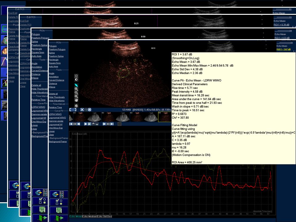
Kvantitativní hodnocení pomocí softwaru (např. QLAB)
")
30
Plocha pod křivkou (AUC) [dB x s] Wash-in analýza [dB/s] Střední tranzitní čas (MTT) [s] Čas do max. nasycení (TTP) [s] 1. Leen E, Gauthier T, Averkiou M, et al. Dynamic contrast enhanced ultrasound assessment of the vascular effects of novel therapeutics in early stage trials. European Radiology. 2012;22(7):1442-1450 2. Bowden DJ, Barrett T. Angiogenesis Imaging in Neoplasia. Journal of Clinical Imaging Science. 2011;1(1):1-7
![ Plocha pod křivkou (AUC) [dB x s] Wash-in analýza [dB/s] Střední tranzitní čas (MTT) [s] Čas do max.](http://images.slideplayer.cz/35/10393428/slides/slide_30.jpg "nasycení (TTP) [s] 1. Leen E, Gauthier T, Averkiou M, et al. Dynamic contrast enhanced ultrasound assessment of the vascular effects of novel therapeutics in early stage trials. European Radiology. 2012;22(7): Bowden DJ, Barrett T. Angiogenesis Imaging in Neoplasia. Journal of Clinical Imaging Science. 2011;1(1):1-7.")
32
PŘED █ Ložisko █ Referenční tkáň

33
PO █ Ložisko █ Referenční tkáň

34
PŘEDPO PředPo Rozdíl Plocha pod křivkou (AUC) [dB x s] 548,14153,07 -73% Wash-in analýza [dB/s] 2,730,54 -80% Střední tranzitní čas (MTT) [s] 23,0014,28 -41% Čas do max. nasycení (TTP) [s] 6,4012,42 +194%
![PŘEDPO PředPo Rozdíl Plocha pod křivkou (AUC) [dB x s] 548,14153,07 -73% Wash-in analýza [dB/s] 2,730,54 -80% Střední tranzitní čas (MTT) [s] 23,0014,28 -41% Čas do max.](http://images.slideplayer.cz/35/10393428/slides/slide_34.jpg "nasycení (TTP) [s] 6,4012, %.")
35
Nízká cena Dobře dostupný, časově nenáročný Bez ionizujícího záření Není nutná speciální příprava pacienta K.l. bez alergických reakcí, minimální kontraindikace Možnost opakování po krátkém čase Možnost kvantitativního hodnocení Bowden DJ, Barrett T. Angiogenesis Imaging in Neoplasia. Journal of Clinical Imaging Science. 2011;1(1):1-7
:1-7.")
36
Závislý na vyšetřujícím lékaři Závislý na pacientovi (obezita, spolupráce) Horší dostupnost u ložisek vysoko pod bránicí Chybí definovaná kritéria v hodnocení léčebné odpovědi 1. Dănilă M, Popescu A, Şirli R, Sporea I, Martie A, Şendroiu M. Contrast enhanced ultrasound (CEUS) in the evaluation of liver metastases. Medical Ultrasonography. 2010;12(3):233-237. 2. Bowden DJ, Barrett T. Angiogenesis Imaging in Neoplasia. Journal of Clinical Imaging Science. 2011;1(1):1-7
 in the evaluation of liver metastases. Medical Ultrasonography. 2010;12(3): Bowden DJ, Barrett T. Angiogenesis Imaging in Neoplasia. Journal of Clinical Imaging Science. 2011;1(1):1-7.")
37
Při antiangiogenní terapii se dlouho nemusí měnit velikost, můžeme ale hodnotit funkční parametry ložisek Kromě PET, SPECT zatím metody experimentální Chybí standardizované protokoly a normy při hodnocení léčebné odpovědi

38
GIST, HCC, meta CRC, Ca prsu a ledvin Nejslibnější metody DWI-MR,CEUS,CTp Hodnocení již od 3-4 dnů Možnost brzy změnit neúčinnou terapii: ◦ benefit pro pacienta, ◦ ekonomický přínos

39
Děkuji, Pane

40
1. Leen E. The role of contrast-enhanced ultrasound in the characterisation of focal liver lesions. European Radiology. 2001;11(SUPPL. 3):E27-E34. 2. Mírka H, Ferda J, Baxa J, et al. Perfuzní CT jater. Čes radiol. 2010;64(4):281-289. 3. Sabir A, Schor-Bardach R, Wilcox CJ, et al. Perfusion MDCT Enables Early Detection of Therapeutic Response to Antiangiogenic Therapy. American Journal of Roentgenology. 2008;191(1):133-139. 4. Pandharipande P, Krinsky G, Rusinek H, Lee V. Perfusion imaging of the liver: Current challenges and future goals. Radiology. 2005;234(3):661-673. 5. Miles K a. Perfusion CT for the assessment of tumour vascularity: Which protocol? British Journal of Radiology. 2003;76(SPEC. ISS. 1):S36-S42. 6. Jiang T, Kambadakone A, Kulkarni N m., Sahani D v. ( 1 ), Zhu A x. ( 3 ). Monitoring response to antiangiogenic treatment and predicting outcomes in advanced hepatocellular carcinoma using image biomarkers, ct perfusion, tumor density, and tumor size (RECIST). Investigative Radiology. 2012;47(1):11-17. 7. Sahani DV, Jiang T, Hayano K, et al. Magnetic resonance imaging biomarkers in hepatocellular carcinoma: association with response and circulating biomarkers after sunitinib therapy. Journal of Hematology & Oncology. 2013;6(1):51. 8. Liver Imaging Research Program | BIRC. 9. Miller JC, Pien HH, Sahani D, Sorensen AG, Thrall JH. Imaging Angiogenesis: Applications and Potential for Drug Development. JNCI: Journal of the National Cancer Institute. 2005;97(3):172-187. 10. How Avastin® (bevacizumab) is different from chemo for rGBM. 11. Sugimoto K, Moriyasu F, Saito K, et al. Hepatocellular carcinoma treated with sorafenib: early detection of treatment response and major adverse events by contrast-enhanced US. Liver International. 2013;33(4):605-615. 12. Goh V, Gourtsoyianni S, Koh D-M. Functional Imaging of the Liver. Seminars in Ultrasound, CT and MRI. 2013;34(1):54-65. 13. Nico B, Benagiano V, Mangieri D, Maruotti N, Vacca A, Ribatti D. Evaluation of microvascular density in tumors: pro and contra. Histology and Histopathology. 2008;23(5):601-607.
:E27-E34. 2. Mírka H, Ferda J, Baxa J, et al. Perfuzní CT jater. Čes radiol. 2010;64(4): 3. Sabir A, Schor-Bardach R, Wilcox CJ, et al. Perfusion MDCT Enables Early Detection of Therapeutic Response to Antiangiogenic Therapy. American Journal of Roentgenology. 2008;191(1): 4. Pandharipande P, Krinsky G, Rusinek H, Lee V. Perfusion imaging of the liver: Current challenges and future goals. Radiology. 2005;234(3): 5. Miles K a. Perfusion CT for the assessment of tumour vascularity: Which protocol. British Journal of Radiology. 2003;76(SPEC. ISS. 1):S36-S42. 6. Jiang T, Kambadakone A, Kulkarni N m., Sahani D v. ( 1 ), Zhu A x. ( 3 ). Monitoring response to antiangiogenic treatment and predicting outcomes in advanced hepatocellular carcinoma using image biomarkers, ct perfusion, tumor density, and tumor size (RECIST). Investigative Radiology. 2012;47(1): 7. Sahani DV, Jiang T, Hayano K, et al. Magnetic resonance imaging biomarkers in hepatocellular carcinoma: association with response and circulating biomarkers after sunitinib therapy. Journal of Hematology & Oncology. 2013;6(1):51. 8. Liver Imaging Research Program | BIRC. 9. Miller JC, Pien HH, Sahani D, Sorensen AG, Thrall JH. Imaging Angiogenesis: Applications and Potential for Drug Development. JNCI: Journal of the National Cancer Institute. 2005;97(3): 10. How Avastin® (bevacizumab) is different from chemo for rGBM. 11. Sugimoto K, Moriyasu F, Saito K, et al. Hepatocellular carcinoma treated with sorafenib: early detection of treatment response and major adverse events by contrast-enhanced US. Liver International. 2013;33(4): 12. Goh V, Gourtsoyianni S, Koh D-M. Functional Imaging of the Liver. Seminars in Ultrasound, CT and MRI. 2013;34(1): 13. Nico B, Benagiano V, Mangieri D, Maruotti N, Vacca A, Ribatti D. Evaluation of microvascular density in tumors: pro and contra. Histology and Histopathology. 2008;23(5):")
41
14. Jiang T, Zhu AX, Sahani DV. Established and novel imaging biomarkers for assessing response to therapy in hepatocellular carcinoma. Journal Of Hepatology. 2013;58(1):169-177. 15. ECR 2012 / C-1677 / Diffusion-weighted whole-body imaging with background body signal suppression (DWIBS): a useful tool in daily practice - EPOS TM. 16. Zocco M, Garcovich M, Lupascu A, et al. Early prediction of response to sorafenib in patients with advanced hepatocellular carcinoma: The role of dynamic contrast enhanced ultrasound. Journal of Hepatology. 2013;59(5):1014-1021. 17. Leen E, Gauthier T, Averkiou M, et al. Dynamic contrast enhanced ultrasound assessment of the vascular effects of novel therapeutics in early stage trials. European Radiology. 2012;22(7):1442-1450. 18. Lim KS. Diffusion-weighted MRI of hepatocellular carcinoma in cirrhosis. Clinical Radiology. 2014;69(1):1-10. 19. Padhani A. Diffusion Magnetic Resonance Imaging in Cancer Patient Management. Seminars in Radiation Oncology. 2011;21(2):119-140. 20. Kim SH, Kamaya A, Willmann JK. CT Perfusion of the Liver: Principles and Applications in Oncology. Radiology. 2014;272(2):322-344. 21. Dănilă M, Popescu A, Şirli R, Sporea I, Martie A, Şendroiu M. Contrast enhanced ultrasound (CEUS) in the evaluation of liver metastases. Medical Ultrasonography. 2010;12(3):233-237. 22. Cui Y, Zhang X-P, Sun Y-S, Tang L, Shen L. Apparent Diffusion Coefficient: Potential Imaging Biomarker for Prediction and Early Detection of Response to Chemotherapy in Hepatic Metastases. Radiology. 2008;248(3):894-900. 23. Bowden DJ, Barrett T. Angiogenesis Imaging in Neoplasia. Journal of Clinical Imaging Science. 2011;1(1):1-7. 24. Lassau N, Chami L, Chebil M, et al. Advanced hepatocellular carcinoma: Early evaluation of response to bevacizumab therapy at dynamic contrast-enhanced us with quantification- preliminary results. Radiology. 2011;258(1):291-300. 25. Heijmen L. Diffusion-weighted MR imaging in liver metastases of colorectal cancer: reproducibility and biological validation. European Radiology. 2013; 23:748–756
: 15. ECR 2012 / C-1677 / Diffusion-weighted whole-body imaging with background body signal suppression (DWIBS): a useful tool in daily practice - EPOS TM. 16. Zocco M, Garcovich M, Lupascu A, et al. Early prediction of response to sorafenib in patients with advanced hepatocellular carcinoma: The role of dynamic contrast enhanced ultrasound. Journal of Hepatology. 2013;59(5): 17. Leen E, Gauthier T, Averkiou M, et al. Dynamic contrast enhanced ultrasound assessment of the vascular effects of novel therapeutics in early stage trials. European Radiology. 2012;22(7): 18. Lim KS. Diffusion-weighted MRI of hepatocellular carcinoma in cirrhosis. Clinical Radiology. 2014;69(1):1-10. 19. Padhani A. Diffusion Magnetic Resonance Imaging in Cancer Patient Management. Seminars in Radiation Oncology. 2011;21(2): 20. Kim SH, Kamaya A, Willmann JK. CT Perfusion of the Liver: Principles and Applications in Oncology. Radiology. 2014;272(2): 21. Dănilă M, Popescu A, Şirli R, Sporea I, Martie A, Şendroiu M. Contrast enhanced ultrasound (CEUS) in the evaluation of liver metastases. Medical Ultrasonography. 2010;12(3): 22. Cui Y, Zhang X-P, Sun Y-S, Tang L, Shen L. Apparent Diffusion Coefficient: Potential Imaging Biomarker for Prediction and Early Detection of Response to Chemotherapy in Hepatic Metastases. Radiology. 2008;248(3): 23. Bowden DJ, Barrett T. Angiogenesis Imaging in Neoplasia. Journal of Clinical Imaging Science. 2011;1(1):1-7. 24. Lassau N, Chami L, Chebil M, et al. Advanced hepatocellular carcinoma: Early evaluation of response to bevacizumab therapy at dynamic contrast-enhanced us with quantification- preliminary results. Radiology. 2011;258(1): 25. Heijmen L. Diffusion-weighted MR imaging in liver metastases of colorectal cancer: reproducibility and biological validation. European Radiology. 2013; 23:748–756.")
Podobné prezentace


>")


