Stáhnout prezentaci
Prezentace se nahrává, počkejte prosím

1
Nové trendy v patologické fyziologii
Chromosomální aberace

2
Dělení buněk mitóza meióza
2 dceřinné buňky s diploidním počtem chromozomů 1 cyklus DNA replikace následuje rozdělení chromozomů a jádra (profáze prometafáze metafáze anafáze telofáze) a násl. celé buňky (cytokineze) meióza 1 cyklus replikace následován 2 cykly segregace chromozomů a buněčného dělení 1. meiotické (redukční) dělení – rozdělení homologních chromozomů významné – odehrává se zde meiotický crossing-over (rekombinace) – žádná z gamet není identická! poruchy rozestupu – např. trisomie 2. meiotické dělení – rozestup sesterských chromatid 2 dceřinné buňky s haploidním počtem chromozomů vznik pohlavních buněk (spermie, vajíčko) dodatečné promíchání genetického materiálu crossing-overem
 a násl. celé buňky (cytokineze) meióza. 1 cyklus replikace následován 2 cykly segregace chromozomů a buněčného dělení. 1. meiotické (redukční) dělení – rozdělení homologních chromozomů. významné – odehrává se zde meiotický crossing-over (rekombinace) – žádná z gamet není identická! poruchy rozestupu – např. trisomie. 2. meiotické dělení – rozestup sesterských chromatid. 2 dceřinné buňky s haploidním počtem chromozomů. vznik pohlavních buněk (spermie, vajíčko) dodatečné promíchání genetického materiálu crossing-overem.")
3
Typy tkání podle regenerační schopnosti
Labilní (intermitotické a postmitotické buňky: kůže, sliznice, hemopoetická tkáň, semenný epitel) Stabilní (reverzibilně postmitotické buňky: Játra-ledviny-pankreas, endotelie, mezoteliální buňky, synoviální krycí buňky, vazivová tkáň, lymfocyty s dlouhým poločasem) Permantní (irreverzibilně postmitotické buňky- gangliové, svalové, vaječné, plasmatické, makrofágy)
 Stabilní (reverzibilně postmitotické buňky: Játra-ledviny-pankreas, endotelie, mezoteliální buňky, synoviální krycí buňky, vazivová tkáň, lymfocyty s dlouhým poločasem) Permantní (irreverzibilně postmitotické buňky- gangliové, svalové, vaječné, plasmatické, makrofágy)")
4
Lidské chromosomy morfologicky barvitelné pouze v průběhu mitózy nebo meiózy, kdy dochází ke kondenzaci v diploidní buňce 23 párů homologních chromosomů (22 párů autosomů a 2 pohlavní chromosomy)
")
5
Karyotyp člověka každý biologický druh má svou charakteristickou chrom. výbavu (počet a morfologii) = karyotyp u člověka mají diploidní bb. 46 chromozomů 22 párů homologních autozomů, 1 pár gomozomů (44XX nebo 44XY) zárodečné (vajíčko, spermie) 23 – haploidní struktura chromozomu centromera telomery (raménka) dlouhé - q krátké – p barvením chromozomů (např. Giems) se dosáhne charakteristického pruhování a tím rozlišení jednotlivých chromozomů
 zárodečné (vajíčko, spermie) 23 – haploidní. struktura chromozomu. centromera. telomery (raménka) dlouhé - q. krátké – p. barvením chromozomů (např. Giems) se dosáhne charakteristického pruhování a tím rozlišení jednotlivých chromozomů.")
6
Karyotyp podle Denverské klasifikace

7
Chromatin chromozom v nedělící se buňce je chromatin rozprostřen volně v jádře u dělící se organizuje do viditelných chromozomů

8
Chromosomové a genové aberace
Chromosomové aberace Strukturní Numerické Genové mutace Vzácné alely Polymorfismy

9
Chromozomální poruchy
aneuploidie (změna počtu chromosomů v sadě) porucha rozdělení sesterských chromozomů [meiotická non-disjunkce] později během rýhování somatická mozaika monosomie gonozomální Turnerův sy. (45, X0) trisomie autozomální Downův sy. (47, XX/XY + 21) Edwardsův sy. (47, XX/XY +18) Patauův sy. (47, XX/XY +13) Klinefelterův sy. (47, XXY) polyploidie (porucha rozdělení celých sad nebo oplození 2 spermiemi [dispermie]) u člověka neslučitelné se životem těhotenství je potraceno molla hydatidosa (a pak těhotenství nutno ukončit potratem) porod novorozence s triploidií – velmi časná letalita
 porucha rozdělení sesterských chromozomů [meiotická non-disjunkce] později během rýhování somatická mozaika. monosomie. gonozomální. Turnerův sy. (45, X0) trisomie. autozomální. Downův sy. (47, XX/XY + 21) Edwardsův sy. (47, XX/XY +18) Patauův sy. (47, XX/XY +13) Klinefelterův sy. (47, XXY) polyploidie (porucha rozdělení celých sad nebo oplození 2 spermiemi [dispermie]) u člověka neslučitelné se životem. těhotenství je potraceno. molla hydatidosa (a pak těhotenství nutno ukončit potratem) porod novorozence s triploidií – velmi časná letalita.")
10
Mozaika Chromosomal mosaicism is when different cells within an individual, who has developed from a single fertilized egg, have a different chromosomal makeup. Most commonly there will be some cells with a typical number of chromosomes (46 chromosomes) and other cells with an altered number or structure of chromosomes. The most common kind of chromosomal mosaicism found at prenatal diagnosis involves trisomy, where the abnormal cells contain 47 chromosomes. Down syndrome mosaicism is an example of trisomy mosaicism. These individuals have some cells with the typical number of chromosomes (46) and some cells with an extra chromosome 21, for a total of 47 chromosomes. Mosaicism may exist for all kinds of chromosome abnormalities (monosomy, triploidy, structural changes, etc). Although more rare, there may even be mosaicism where both different cell types are abnormal in structure or number, and there are no normal cells involved. How does mosaicism occur? The cells with abnormal chromosomes may be found in mutiple tissues, or in just one tissue. Consider the example of trisomy 21 mosaicism or Down syndrome mosaicism. In one individual, if we look at a blood sample we may find some cells with 47 chromosomes and some with 46 chromosomes. If we look at a skin sample we may also see some cells with the abnormal number of chromosomes (47) and some with the normal number of chromosomes (46). This is illustrated in diagram a. On the other hand if we look at another individual, it is possible that in one tissue, say skin, we see mostly cells with 46 chromosomes, but in another tissue, say the intestine, we may see mostly cells with an extra chromosome 21, for a total of 47 chromosomes. This is illustrated in diagram b.
 and other cells with an altered number or structure of chromosomes. The most common kind of chromosomal mosaicism found at prenatal diagnosis involves trisomy, where the abnormal cells contain 47 chromosomes. Down syndrome mosaicism is an example of trisomy mosaicism. These individuals have some cells with the typical number of chromosomes (46) and some cells with an extra chromosome 21, for a total of 47 chromosomes. Mosaicism may exist for all kinds of chromosome abnormalities (monosomy, triploidy, structural changes, etc). Although more rare, there may even be mosaicism where both different cell types are abnormal in structure or number, and there are no normal cells involved. How does mosaicism occur The cells with abnormal chromosomes may be found in mutiple tissues, or in just one tissue. Consider the example of trisomy 21 mosaicism or Down syndrome mosaicism. In one individual, if we look at a blood sample we may find some cells with 47 chromosomes and some with 46 chromosomes. If we look at a skin sample we may also see some cells with the abnormal number of chromosomes (47) and some with the normal number of chromosomes (46). This is illustrated in diagram a. On the other hand if we look at another individual, it is possible that in one tissue, say skin, we see mostly cells with 46 chromosomes, but in another tissue, say the intestine, we may see mostly cells with an extra chromosome 21, for a total of 47 chromosomes. This is illustrated in diagram b.")
11
How does trisomy mosaicism occur?
All of the cells in our body come from a single cell, the fertilized egg or zygote. In order for the zygote to develop into a baby, this single cell must grow and divide. Before cells can divide each chromosome must make an identical copy of itself. At cell division each chromosome and its identical copy pull apart into two separate cells. Now the resulting cells, also called daughter cells, have the same chromosome make-up as the original cell. The two new cells will repeat this process. In each cell, the chromosomes will duplicate and divide into two new cells. The result, is 4 cells identical to the first original cell. This process of cell division is called mitosis. Sometimes a mistake can occur when the chromosomes are separating into the two daughter cells. An extra chromosome may travel into the wrong cell or a chromosome may get lost in the separation of the cells. The result would be two daughter cells with different chromosomal make-up.

13
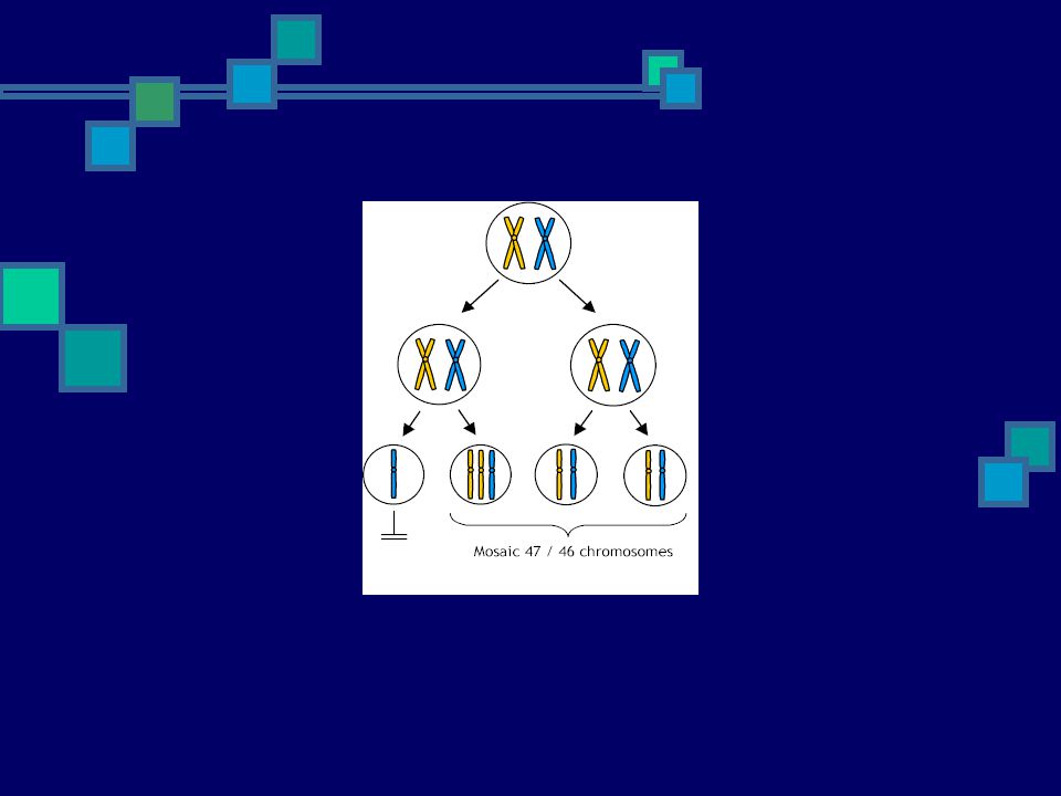
Trisomy mosaicism can occur in one of two ways:
In an abnormal fertilized egg with 47 chromosomes, one of the cells may lose the extra chromosome at cell division, leaving 46 chromosomes in that cell. All cells that are derived from that cell will have 46 chromosomes. The rest of the cells will have 47 chromosomes. In a typical zygote with 46 chromosomes, at cell division one of the cells may retain a duplicated copy of one of the chromosomes. This produces a cell with 47 chromosomes. All cells that are derived from that cell also have 47 chromosomes. The rest of the cells will have 46 chromosomes. In both cases the result is a baby with two different cell lines, one cell line with 46 chromosomes and one with 47 chromosomes.

14
Trisomy mosaicism Trisomy mosaicism can originate in two ways.
A. Somatic origin Mitotic non-disjunction in a cell of a fertilized egg with the typical 46 chromosomes, leads to a different cell line with an additional chromosome. The cell with three copies of the chromosome may continue to grow, however the cell with only one copy of the chromosome is more often severely disadvantaged and usually will not continue to reproduce (Gardner & Sutherland, 1996). .
. .")
16
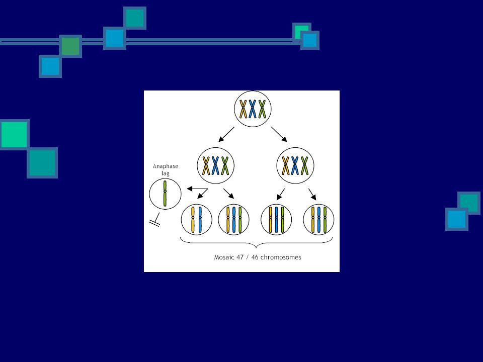
Trisomy mosaicism B. Meiotic origin
The other mechanism, which involves loss of the extra chromosome, can occur through a process called anaphase lag in an abnormal fertilized egg with 47 chromosomes. In the process of anaphase lag, the extra chromosome fails to be included in the formation of the new cell and becomes isolated and eventually lost.

18
The mosaic pattern depends on many factors.
1. Number of cells present at the time of the non-disjunction mistake A very early mistake will effect a greater proportion of the cells in the baby. Mosaicism originating from an early mistake, either in the first or second division of the fertilized egg, leads to generalized mosaicism, since most tissues of the baby are affected, often in a "patchy" way. A mistake which occurs at a later stage, for example at the 64-celled blastocyst stage, will effect a smaller proportion of the cells in the baby. "Later errors" may lead to an abnormal line of cells confined to a certain area or tissue in the developing individual.

19
The mosaic pattern depends on many factors.
Theoretically, if the mistake happens just in cells that are destined to become the placenta then the abnormal cells may be confined to the placenta and may not be found in the baby. Or if trisomic rescue occurs in the cells that are destined to become the baby, then the abnormal cells may be confined to the placenta and not found in the baby. This is called (mozaika omezená na placentu) confined placental mosaicism. If the mistake happens just in cells that are destined to become the baby, then the abnormal cells will be confined to the baby. This is called (mozaika omezená na embryo) confined embryonic mosaicism. Many more cells contribute to the placenta. It is extremely difficult to diagnose confined mosaicism with certainty because it is impossible to sample all tissues in an individual.
 confined placental mosaicism. If the mistake happens just in cells that are destined to become the baby, then the abnormal cells will be confined to the baby. This is called (mozaika omezená na embryo) confined embryonic mosaicism. Many more cells contribute to the placenta. It is extremely difficult to diagnose confined mosaicism with certainty because it is impossible to sample all tissues in an individual.")
20
The mosaic pattern depends on many factors.
2. Type of cells involved The development and health of the affected baby also depends on the type of cells affected by the mistake. The change in number of chromosomes is only important if it affects the function of the tissue(s) involved. If the duplicated chromosome contains genetic instructions that are crucial to the function of that tissue, the effect on the overall function of the tissue might be impaired or, on the other hand, there may even be selection against the affected cells. 3. Survival of trisomic cells Also important in determining the outcome is the ability of the abnormal cells to survive. The question is, can the cells with the chromosome mistake continue to reproduce? Certain mechanisms involved in cell selection may prevent the abnormal trisomic cells from reproducing, thus minimizing or eliminating the effect of the original non-disjunction error. The specific chromosome involved seems to play a role in determining the survival of the cells. Studies of cell cultures suggest that trisomic cells generally divide less quickly and undergo cell death more commonly than diploid cells.
 involved. If the duplicated chromosome contains genetic instructions that are crucial to the function of that tissue, the effect on the overall function of the tissue might be impaired or, on the other hand, there may even be selection against the affected cells. 3. Survival of trisomic cells. Also important in determining the outcome is the ability of the abnormal cells to survive. The question is, can the cells with the chromosome mistake continue to reproduce Certain mechanisms involved in cell selection may prevent the abnormal trisomic cells from reproducing, thus minimizing or eliminating the effect of the original non-disjunction error. The specific chromosome involved seems to play a role in determining the survival of the cells. Studies of cell cultures suggest that trisomic cells generally divide less quickly and undergo cell death more commonly than diploid cells.")
21
Jak se diagnostikuje chromosomální mozaika?
Během prenatální diagnostiky Z krve nebo kožní biopsie Během preimplantační diagnostiky

22
Klinický dopad mozaiky
When chromosomal mosaicism arises during development, pregnancy outcome depends on which tissue, and how much of that tissue is abnormal. In theory, cases with a relatively high proportion of trisomic cells are more likely to be associated with an abnormal outcome than those with a low proportion of trisomic cells. That is, if a majority of the cells are abnormal then human development is likely to be abnormal. If only a tiny fraction of some tissue were involved, the aneuploidy would likely have little effect on growth and development. Perhaps many people carry a tiny and completely unimportant abnormal cell line somewhere in their body. However, a very minor degree of mosaicism could still be important if a crucial tissue carries the abnormal cells. For example, an abnormal chromosome change confined to one part of the brain could theoretically impair neurological function (Gardner & Sutherland, 1996).
.")
23
Klinický dopad mozaiky
As a general principle, an individual with a chromosome abnormality in only some of their tissues is likely to have less severe but qualitatively similar clinical features to that of someone with the non-mosaic form of the same chromosome abnormality. For example mosaic Down syndrome can be associated with a less characteristic facial appearance and milder mental impairment than the those with typical trisomy 21. Some chromosome changes can only exist in a mosaic form, because in a non-mosaic form they are lethal. Sometimes if the distribution of the aneuploid cell line is asymetric, the body shape or appearance may be asymmetric. Generally it is the cells that are aneuploid that are smaller and less developed (Gardner & Sutherland, 1996). It is worth noting though that chromosomally abnormal cells may also arise with age and contribute to such health problems as the occurrence of cancer. However, most age-related chromosome changes are likely either eliminated due to poor cell growth or have no obvious harmful effect. For example, 45, X0 cells are increasingly common in female blood cells as they age, but appear to have no harmful effect.
. It is worth noting though that chromosomally abnormal cells may also arise with age and contribute to such health problems as the occurrence of cancer. However, most age-related chromosome changes are likely either eliminated due to poor cell growth or have no obvious harmful effect. For example, 45, X0 cells are increasingly common in female blood cells as they age, but appear to have no harmful effect.")
24
Děkuji za pozornost!

Podobné prezentace