Stáhnout prezentaci
Prezentace se nahrává, počkejte prosím

1
Biofyzika vnímání světelných podnětů
Přednášky z lékařské biofyziky Masarykova universita v Brně Biofyzikální ústav Biofyzika vnímání světelných podnětů

2
Obsah přednášky Základní vlastnosti světla Anatomie oka
Optické vlastnosti oka Sítnice – biologický detektor světla Barevné vidění

3
Základní vlastnosti světla
Viditelné elektromagnetické záření: λ = 380 – 760 nm Kratší vlnové délky – Ultrafialové světlo (UV) Delší vlnové délky – Infračervené světlo (IR) Viditelné světlo – (VIS) Prostředí, kterým se světlo šíří, se nazývá optické prostředí. V homogenním prostředí se světlo šíří přímočaře, kolmo k vlnoplochám, čáry směru šíření se označují jako světelné paprsky. Rychlost světla (ve vakuu) c = ms-1 ≈ ms-1
 Delší vlnové délky – Infračervené světlo (IR) Viditelné světlo – (VIS) Prostředí, kterým se světlo šíří, se nazývá optické prostředí. V homogenním prostředí se světlo šíří přímočaře, kolmo k vlnoplochám, čáry směru šíření se označují jako světelné paprsky. Rychlost světla (ve vakuu) c = ms-1 ≈ ms-1.")
4
Zdroje viditelného (VIS) světla
Přirozené Umělé Přirozené: Slunce Slunce je zdrojem energie pro život a nelze si představit život bez něj. Umělé: žárovky, fluorescenční zdroje, laser…

5
Polychromatické a monochromatické světlo, koherence
Polychromatické či bílé světlo je světlem o různých vlnových délkách. Monochromatické světlo je světlem o jediné vlnové délce. Dle svého „fázového“ charakteru může být světlo Koherentní - Koherentní světlo jsou světelné vlny jsoucí vzájemně „ve fázi“, tj. mají stejnou fázi v též vzdálenosti od zdroje. Světlo emitované laserem je koherentní. Nekoherentní - Světelné vlny nejsou vzájemně „ve fázi“. Světlo žárovek nebo Slunce je nekoherentní.

6
Odraz a lom světla Odraz – zákon odrazu: Úhel odrazu ’ je roven úhlu dopadu. Odražený paprsek se šíří v rovině dopadu. Lom: Když světlo prochází z jednoho prostředí do druhého, jeho svazek mění směr na rozhraní mezi dvěma prostředími. Tato vlastnost optického prostředí je popsána indexem lomu n = c/v [ bezrozměrová veličina] n – index lomu prostředí c – rychlost světla ve vakuu v – rychlost světla v prostředí index lomu vakua je 1

7
Odraz a lom světla Snellův zákon (zákon lomu) α – úhel dopadu (v prostředí 1) β – úhel lomu (v prostředí 2) (úhly měříme od kolmice!) n1, n2 – indexy lomu v1, v2, – rychlosti světla v daných prostředích n je velká: velká optická hustota) n je malél: malá optická hustota n1 > n2 – dochází k lomu od kolmice n1 < n2 – dochází k lomu ke kolmici
 α – úhel dopadu (v prostředí 1) β – úhel lomu (v prostředí 2) (úhly měříme od kolmice!) n1, n2 – indexy lomu v1, v2, – rychlosti světla v daných prostředích n je velká: velká optická hustota) n je malél: malá optická hustota n1 > n2 – dochází k lomu od kolmice n1 < n2 – dochází k lomu ke kolmici.")
8
Rovnice „brusičů čoček“
f – ohnisková vzdálenost [m] n2 – index lomu čočky n1 – index lomu okolního prostředí r1, r2 – poloměry křivosti čočky

9
Obecné principy optického zobrazení
Skutečný obraz (lze promítnout): sbíhavost paprsků Neskutečný obraz (nelze promítnout): rozbíhavost paprsků Optická osa – osa centrovaného systému optických rozhraní Ohnisko - bod, v němž se protínají paprsky dopadající na čočku (zakřivené zrcadlo) rovnoběžně s optickou osou. Rozlišujeme přední (předmětové) ohnisko a zadní (obrazové) ohnisko Ohnisková vzdálenost f [m] je vzdálenost ohniska od středu čočky nebo zrcadla Poloměry křivosti jsou kladné (záporné), pokud je příslušná čočka nebo povrch zrcadla konvexní (konkávní). Optická mohutnost (síla čočky): převrácená hodnota ohniskové vzdálenosti = D = S = 1/f [m-1 = dpt = D (dioptrie)] Spojné čočky: f a jsou kladné Rozptylné čočky: f a jsou záporné
: sbíhavost paprsků. Neskutečný obraz (nelze promítnout): rozbíhavost paprsků. Optická osa – osa centrovaného systému optických rozhraní. Ohnisko - bod, v němž se protínají paprsky dopadající na čočku (zakřivené zrcadlo) rovnoběžně s optickou osou. Rozlišujeme přední (předmětové) ohnisko a zadní (obrazové) ohnisko. Ohnisková vzdálenost f [m] je vzdálenost ohniska od středu čočky nebo zrcadla. Poloměry křivosti jsou kladné (záporné), pokud je příslušná čočka nebo povrch zrcadla konvexní (konkávní). Optická mohutnost (síla čočky): převrácená hodnota ohniskové vzdálenosti. = D = S = 1/f [m-1 = dpt = D (dioptrie)] Spojné čočky: f a jsou kladné. Rozptylné čočky: f a jsou záporné.")
10
Rovnice čočky Paprsky rovnoběžné s optickou osou se lámou do obrazového ohniska (u spojné čočky), nebo tak, že se zdají vycházet z předmětového ohniska (u rozptylky). Směr paprsků procházejících středem čočky se nemění. Rovnice čočky: a – vzdálenost předmětu [m] b – vzdálenost obrazu [m] Znaménková konvence: a je kladné před čočkou, záporné za čočkou; b je záporné před čočkou (obraz je neskutečný), kladné za čočkou (obraz je skutečný)
, nebo tak, že se zdají vycházet z předmětového ohniska (u rozptylky). Směr paprsků procházejících středem čočky se nemění. Rovnice čočky: a – vzdálenost předmětu [m] b – vzdálenost obrazu [m] Znaménková konvence: a je kladné před čočkou, záporné za čočkou; b je záporné před čočkou (obraz je neskutečný), kladné za čočkou (obraz je skutečný)")
11
Viditelné spektrum Lidské oko je citlivé na světlo od vlnové délky zhruba 380 nm (fialové) do asi 760 nm (červené). Náš zrakový analyzátor vnímá tento rozsah vlnových délek jako spojité duhové spektrum. Nazýváme je viditelné spektrum. Viz obrázek.
 do asi 760 nm (červené). Náš zrakový analyzátor vnímá tento rozsah vlnových délek jako spojité duhové spektrum. Nazýváme je viditelné spektrum. Viz obrázek.")
12
Anatomie oka

13
Jak pracuje lidské oko? Jednotlivé části oka pracují podobným způsobem jako části fotografického přístroje. Každá část je důležitá pro zřetelné vidění. How Does The Human Eye Work? The individual components of the eye work in a manner similar to a camera. Each part plays a vital role in providing clear vision. So think of the eye as a camera with the cornea, behaving much like a lens cover. As the eye's main focusing element, the cornea takes widely diverging rays of light and bends them through the pupil, the dark, round opening in the center of the colored iris. The iris and pupil act like the aperture of a camera. Next in line is the lens which acts like the lens in a camera, helping to focus light to the back of the eye. Note that the lens is the part which becomes cloudy and is removed during cataract surgery to be replaced by an artificial implant nowadays. The Camera The Human Eye The very back of the eye is lined with a layer called the retina which acts very much like the film of the camera. The retina is a membrane containing photoreceptor nerve cells that lines the inside back wall of the eye. The photoreceptor nerve cells of the retina change the light rays into electrical impulses and send them through the optic nerve to the brain where an image is perceived. The center 10% of the retina is called the macula. This is responsible for your sharp vision, your reading vision. The peripheral retina is responsible for the peripheral vision. As with the camera, if the "film" is bad in the eye (i.e. the retina), no matter how good the rest of the eye is, you will not get a good picture. The human eye is remarkable. It accommodates to changing lighting conditions and focuses light rays originating from various distances from the eye. When all of the components of the eye function properly, light is converted to impulses and conveyed to the brain where an image is perceived. Glossary of Eye Terms: Anterior Chamber The cavity in the front part of the eye between the lens and cornea is called the Anterior Chamber. It is filled with Aqueous, a water-like fluid. This fluid is produced by the ciliary body and drains back into the blood circulation through channels in the chamber angle. It is turned over every100 minutes. Chamber Angle Located at the junction of the cornea, iris, and sclera, the anterior chamber angle extends 360 degrees at the perimeter of the iris. Channels here allow aqueous fluid to drain back into the blood circulation from the eye. May be obstructed in glaucoma. Ciliary Body A structure located behind the iris (rarely visible) which produces aqueous fluid that fills the front part of the eye and thus maintains the eye pressure. It also allows focusing of the lens. Conjunctiva A thin lining over the sclera, or white part of the eye. This also lines the inside of the eyelids. Cell in the conjunctiva produce mucous, which helps to lubricate the eye. Cornea The transparent, outer "window" and primary focusing element of the eye. The outer layer of the cornea is known as epithelium. Its main job is to protect the eye. The epithelium is made up of transparent cells that have the ability to regenerate quickly. The inner layer of the cornea is also made up of transparent tissue, which allows light to pass. Hyaloid Canal A narrow channel that runs from the optic disc to the back surface of the lens. It serves an embryologic function prior to birth but none afterwards. Iris Inside the anterior chamber is the iris. This is the part of the eye which is responsible for one's eye color. It acts like the diaphragm of a camera, dilating and constricting the pupil to allow more or less light into the eye. Pupil The dark opening in the center of the colored iris that controls how much light enters the eye. The colored iris functions like the iris of a camera, opening and closing, to control the amount of light entering through the pupil. Lens The part of the eye immediately behind the iris that performs delicate focusing of light rays upon the retina. In persons under 40, the lens is soft and pliable, allowing for fine focusing from a wide variety of distances. For individuals over 40, the lens begins to become less pliable, making focusing upon objects near to the eye more difficult. This is known as presbyopia. Macula The part of the retina which is most sensitive, and is responsible for the central (or reading) vision. It is located near the optic nerve directly at the back of the eye (on the inside). This area is also responsible for color vision. Optic Disc The position in the back of the eye where the nerve (along with an artery and vein) enters the eye corresponds to the "blind spot" since there are no rods or cones in these location. Normally, a person does not notice this blind spot since rapid movements of the eye and processing in the brain compensate for this absent information. This is the area that the ophthalmologist studies when evaluating a patient for glaucoma, a condition where the optic nerve becomes damaged often due to high pressure within the eye. As it looks like a cup when viewed with an ophthalmoscope, it is sometimes referred to as the Optic Cup. Optic Nerve The optic nerve is the structure which takes the information from the retina as electrical signals and delivers it to the brain where this information is interpreted as a visual image. The optic nerve consists of a bundle of about one million nerve fibers. Retina The membrane lining the back of the eye that contains photoreceptor cells. These photoreceptor nerve cells react to the presence and intensity of light by sending an impulse to the brain via the optic nerve. In the brain, the multitude of nerve impulses received from the photoreceptor cells in the retina are assimilated into an image. Sclera The white, tough wall of the eye. Few diseases affect this layer. It is covered by the episclera (a fibrous layer between the conjunctiva and sclera ) and conjunctiva, and eye muscles are connected to this. Vitreous Next in our voyage through the eye is the vitreous. This is a jelly-like substance that fills the body of the eye. It is normally clear. In early life, it is firmly attached to the retina behind it. With age, the vitreous becomes more water-like and may detach from the retina. Often, little clumps or strands of the jelly form and cast shadows which are perceived as "floaters". While frequently benign, sometimes floaters can be a sign of a more serious condition such as a retinal tear or detachment and should be investigated with a thorough ophthalmologic examination. How Does The Human Eye Work? The individual components of the eye work in a manner similar to a camera. Each part plays a vital role in providing clear vision. So think of the eye as a camera with the cornea, behaving much like a lens cover. As the eye's main focusing element, the cornea takes widely diverging rays of light and bends them through the pupil, the dark, round opening in the center of the colored iris. The iris and pupil act like the aperture of a camera. Next in line is the lens which acts like the lens in a camera, helping to focus light to the back of the eye. Note that the lens is the part which becomes cloudy and is removed during cataract surgery to be replaced by an artificial implant nowadays. Anterior Chamber The cavity in the front part of the eye between the lens and cornea is called the Anterior Chamber. It is filled with Aqueous, a water-like fluid. This fluid is produced by the ciliary body and drains back into the blood circulation through channels in the chamber angle. It is turned over every100 minutes. Chamber Angle Located at the junction of the cornea, iris, and sclera, the anterior chamber angle extends 360 degrees at the perimeter of the iris. Channels here allow aqueous fluid to drain back into the blood circulation from the eye. May be obstructed in glaucoma. Ciliary Body A structure located behind the iris (rarely visible) which produces aqueous fluid that fills the front part of the eye and thus maintains the eye pressure. It also allows focusing of the lens. Conjunctiva A thin lining over the sclera, or white part of the eye. This also lines the inside of the eyelids. Cell in the conjunctiva produce mucous, which helps to lubricate the eye. Cornea The transparent, outer "window" and primary focusing element of the eye. The outer layer of the cornea is known as epithelium. Its main job is to protect the eye. The epithelium is made up of transparent cells that have the ability to regenerate quickly. The inner layer of the cornea is also made up of transparent tissue, which allows light to pass. Hyaloid Canal A narrow channel that runs from the optic disc to the back surface of the lens. It serves an embryologic function prior to birth but none afterwards. Iris Inside the anterior chamber is the iris. This is the part of the eye which is responsible for one's eye color. It acts like the diaphragm of a camera, dilating and constricting the pupil to allow more or less light into the eye. Pupil The dark opening in the center of the colored iris that controls how much light enters the eye. The colored iris functions like the iris of a camera, opening and closing, to control the amount of light entering through the pupil. Lens The part of the eye immediately behind the iris that performs delicate focusing of light rays upon the retina. In persons under 40, the lens is soft and pliable, allowing for fine focusing from a wide variety of distances. For individuals over 40, the lens begins to become less pliable, making focusing upon objects near to the eye more difficult. This is known as presbyopia. Macula The part of the retina which is most sensitive, and is responsible for the central (or reading) vision. It is located near the optic nerve directly at the back of the eye (on the inside). This area is also responsible for color vision. Optic Disc The position in the back of the eye where the nerve (along with an artery and vein) enters the eye corresponds to the "blind spot" since there are no rods or cones in these location. Normally, a person does not notice this blind spot since rapid movements of the eye and processing in the brain compensate for this absent information. This is the area that the ophthalmologist studies when evaluating a patient for glaucoma, a condition where the optic nerve becomes damaged often due to high pressure within the eye. As it looks like a cup when viewed with an ophthalmoscope, it is sometimes referred to as the Optic Cup. Optic Nerve The optic nerve is the structure which takes the information from the retina as electrical signals and delivers it to the brain where this information is interpreted as a visual image. The optic nerve consists of a bundle of about one million nerve fibers. Retina The membrane lining the back of the eye that contains photoreceptor cells. These photoreceptor nerve cells react to the presence and intensity of light by sending an impulse to the brain via the optic nerve. In the brain, the multitude of nerve impulses received from the photoreceptor cells in the retina are assimilated into an image. Sclera The white, tough wall of the eye. Few diseases affect this layer. It is covered by the episclera (a fibrous layer between the conjunctiva and sclera ) and conjunctiva, and eye muscles are connected to this. Vitreous Next in our voyage through the eye is the vitreous. This is a jelly-like substance that fills the body of the eye. It is normally clear. In early life, it is firmly attached to the retina behind it. With age, the vitreous becomes more water-like and may detach from the retina. Often, little clumps or strands of the jelly form and cast shadows which are perceived as "floaters". While frequently benign, sometimes floaters can be a sign of a more serious condition such as a retinal tear or detachment and should be investigated with a thorough ophthalmologic examination. How Does The Human Eye Work? The individual components of the eye work in a manner similar to a camera. Each part plays a vital role in providing clear vision. So think of the eye as a camera with the cornea, behaving much like a lens cover. As the eye's main focusing element, the cornea takes widely diverging rays of light and bends them through the pupil, the dark, round opening in the center of the colored iris. The iris and pupil act like the aperture of a camera. Next in line is the lens which acts like the lens in a camera, helping to focus light to the back of the eye. Note that the lens is the part which becomes cloudy and is removed during cataract surgery to be replaced by an artificial implant nowadays. Anterior Chamber The cavity in the front part of the eye between the lens and cornea is called the Anterior Chamber. It is filled with Aqueous, a water-like fluid. This fluid is produced by the ciliary body and drains back into the blood circulation through channels in the chamber angle. It is turned over every100 minutes. Chamber Angle Located at the junction of the cornea, iris, and sclera, the anterior chamber angle extends 360 degrees at the perimeter of the iris. Channels here allow aqueous fluid to drain back into the blood circulation from the eye. May be obstructed in glaucoma. Ciliary Body A structure located behind the iris (rarely visible) which produces aqueous fluid that fills the front part of the eye and thus maintains the eye pressure. It also allows focusing of the lens. Conjunctiva A thin lining over the sclera, or white part of the eye. This also lines the inside of the eyelids. Cell in the conjunctiva produce mucous, which helps to lubricate the eye. Cornea The transparent, outer "window" and primary focusing element of the eye. The outer layer of the cornea is known as epithelium. Its main job is to protect the eye. The epithelium is made up of transparent cells that have the ability to regenerate quickly. The inner layer of the cornea is also made up of transparent tissue, which allows light to pass. Hyaloid Canal A narrow channel that runs from the optic disc to the back surface of the lens. It serves an embryologic function prior to birth but none afterwards. Iris Inside the anterior chamber is the iris. This is the part of the eye which is responsible for one's eye color. It acts like the diaphragm of a camera, dilating and constricting the pupil to allow more or less light into the eye. Pupil The dark opening in the center of the colored iris that controls how much light enters the eye. The colored iris functions like the iris of a camera, opening and closing, to control the amount of light entering through the pupil. Lens The part of the eye immediately behind the iris that performs delicate focusing of light rays upon the retina. In persons under 40, the lens is soft and pliable, allowing for fine focusing from a wide variety of distances. For individuals over 40, the lens begins to become less pliable, making focusing upon objects near to the eye more difficult. This is known as presbyopia. Macula The part of the retina which is most sensitive, and is responsible for the central (or reading) vision. It is located near the optic nerve directly at the back of the eye (on the inside). This area is also responsible for color vision. Optic Disc The position in the back of the eye where the nerve (along with an artery and vein) enters the eye corresponds to the "blind spot" since there are no rods or cones in these location. Normally, a person does not notice this blind spot since rapid movements of the eye and processing in the brain compensate for this absent information. This is the area that the ophthalmologist studies when evaluating a patient for glaucoma, a condition where the optic nerve becomes damaged often due to high pressure within the eye. As it looks like a cup when viewed with an ophthalmoscope, it is sometimes referred to as the Optic Cup. Optic Nerve The optic nerve is the structure which takes the information from the retina as electrical signals and delivers it to the brain where this information is interpreted as a visual image. The optic nerve consists of a bundle of about one million nerve fibers. Retina The membrane lining the back of the eye that contains photoreceptor cells. These photoreceptor nerve cells react to the presence and intensity of light by sending an impulse to the brain via the optic nerve. In the brain, the multitude of nerve impulses received from the photoreceptor cells in the retina are assimilated into an image. Sclera The white, tough wall of the eye. Few diseases affect this layer. It is covered by the episclera (a fibrous layer between the conjunctiva and sclera ) and conjunctiva, and eye muscles are connected to this. Vitreous Next in our voyage through the eye is the vitreous. This is a jelly-like substance that fills the body of the eye. It is normally clear. In early life, it is firmly attached to the retina behind it. With age, the vitreous becomes more water-like and may detach from the retina. Often, little clumps or strands of the jelly form and cast shadows which are perceived as "floaters". While frequently benign, sometimes floaters can be a sign of a more serious condition such as a retinal tear or detachment and should be investigated with a thorough ophthalmologic examination. Fot. přístroj Lidské oko
, no matter how good the rest of the eye is, you will not get a good picture. The human eye is remarkable. It accommodates to changing lighting conditions and focuses light rays originating from various distances from the eye. When all of the components of the eye function properly, light is converted to impulses and conveyed to the brain where an image is perceived. Glossary of Eye Terms: Anterior Chamber The cavity in the front part of the eye between the lens and cornea is called the Anterior Chamber. It is filled with Aqueous, a water-like fluid. This fluid is produced by the ciliary body and drains back into the blood circulation through channels in the chamber angle. It is turned over every100 minutes. Chamber Angle Located at the junction of the cornea, iris, and sclera, the anterior chamber angle extends 360 degrees at the perimeter of the iris. Channels here allow aqueous fluid to drain back into the blood circulation from the eye. May be obstructed in glaucoma. Ciliary Body A structure located behind the iris (rarely visible) which produces aqueous fluid that fills the front part of the eye and thus maintains the eye pressure. It also allows focusing of the lens. Conjunctiva A thin lining over the sclera, or white part of the eye. This also lines the inside of the eyelids. Cell in the conjunctiva produce mucous, which helps to lubricate the eye. Cornea The transparent, outer window and primary focusing element of the eye. The outer layer of the cornea is known as epithelium. Its main job is to protect the eye. The epithelium is made up of transparent cells that have the ability to regenerate quickly. The inner layer of the cornea is also made up of transparent tissue, which allows light to pass. Hyaloid Canal A narrow channel that runs from the optic disc to the back surface of the lens. It serves an embryologic function prior to birth but none afterwards. Iris Inside the anterior chamber is the iris. This is the part of the eye which is responsible for one s eye color. It acts like the diaphragm of a camera, dilating and constricting the pupil to allow more or less light into the eye. Pupil The dark opening in the center of the colored iris that controls how much light enters the eye. The colored iris functions like the iris of a camera, opening and closing, to control the amount of light entering through the pupil. Lens The part of the eye immediately behind the iris that performs delicate focusing of light rays upon the retina. In persons under 40, the lens is soft and pliable, allowing for fine focusing from a wide variety of distances. For individuals over 40, the lens begins to become less pliable, making focusing upon objects near to the eye more difficult. This is known as presbyopia. Macula The part of the retina which is most sensitive, and is responsible for the central (or reading) vision. It is located near the optic nerve directly at the back of the eye (on the inside). This area is also responsible for color vision. Optic Disc The position in the back of the eye where the nerve (along with an artery and vein) enters the eye corresponds to the blind spot since there are no rods or cones in these location. Normally, a person does not notice this blind spot since rapid movements of the eye and processing in the brain compensate for this absent information. This is the area that the ophthalmologist studies when evaluating a patient for glaucoma, a condition where the optic nerve becomes damaged often due to high pressure within the eye. As it looks like a cup when viewed with an ophthalmoscope, it is sometimes referred to as the Optic Cup. Optic Nerve The optic nerve is the structure which takes the information from the retina as electrical signals and delivers it to the brain where this information is interpreted as a visual image. The optic nerve consists of a bundle of about one million nerve fibers. Retina The membrane lining the back of the eye that contains photoreceptor cells. These photoreceptor nerve cells react to the presence and intensity of light by sending an impulse to the brain via the optic nerve. In the brain, the multitude of nerve impulses received from the photoreceptor cells in the retina are assimilated into an image. Sclera The white, tough wall of the eye. Few diseases affect this layer. It is covered by the episclera (a fibrous layer between the conjunctiva and sclera ) and conjunctiva, and eye muscles are connected to this. Vitreous Next in our voyage through the eye is the vitreous. This is a jelly-like substance that fills the body of the eye. It is normally clear. In early life, it is firmly attached to the retina behind it. With age, the vitreous becomes more water-like and may detach from the retina. Often, little clumps or strands of the jelly form and cast shadows which are perceived as floaters . While frequently benign, sometimes floaters can be a sign of a more serious condition such as a retinal tear or detachment and should be investigated with a thorough ophthalmologic examination. How Does The Human Eye Work The individual components of the eye work in a manner similar to a camera. Each part plays a vital role in providing clear vision. So think of the eye as a camera with the cornea, behaving much like a lens cover. As the eye s main focusing element, the cornea takes widely diverging rays of light and bends them through the pupil, the dark, round opening in the center of the colored iris. The iris and pupil act like the aperture of a camera. Next in line is the lens which acts like the lens in a camera, helping to focus light to the back of the eye. Note that the lens is the part which becomes cloudy and is removed during cataract surgery to be replaced by an artificial implant nowadays. Anterior Chamber The cavity in the front part of the eye between the lens and cornea is called the Anterior Chamber. It is filled with Aqueous, a water-like fluid. This fluid is produced by the ciliary body and drains back into the blood circulation through channels in the chamber angle. It is turned over every100 minutes. Chamber Angle Located at the junction of the cornea, iris, and sclera, the anterior chamber angle extends 360 degrees at the perimeter of the iris. Channels here allow aqueous fluid to drain back into the blood circulation from the eye. May be obstructed in glaucoma. Ciliary Body A structure located behind the iris (rarely visible) which produces aqueous fluid that fills the front part of the eye and thus maintains the eye pressure. It also allows focusing of the lens. Conjunctiva A thin lining over the sclera, or white part of the eye. This also lines the inside of the eyelids. Cell in the conjunctiva produce mucous, which helps to lubricate the eye. Cornea The transparent, outer window and primary focusing element of the eye. The outer layer of the cornea is known as epithelium. Its main job is to protect the eye. The epithelium is made up of transparent cells that have the ability to regenerate quickly. The inner layer of the cornea is also made up of transparent tissue, which allows light to pass. Hyaloid Canal A narrow channel that runs from the optic disc to the back surface of the lens. It serves an embryologic function prior to birth but none afterwards. Iris Inside the anterior chamber is the iris. This is the part of the eye which is responsible for one s eye color. It acts like the diaphragm of a camera, dilating and constricting the pupil to allow more or less light into the eye. Pupil The dark opening in the center of the colored iris that controls how much light enters the eye. The colored iris functions like the iris of a camera, opening and closing, to control the amount of light entering through the pupil. Lens The part of the eye immediately behind the iris that performs delicate focusing of light rays upon the retina. In persons under 40, the lens is soft and pliable, allowing for fine focusing from a wide variety of distances. For individuals over 40, the lens begins to become less pliable, making focusing upon objects near to the eye more difficult. This is known as presbyopia. Macula The part of the retina which is most sensitive, and is responsible for the central (or reading) vision. It is located near the optic nerve directly at the back of the eye (on the inside). This area is also responsible for color vision. Optic Disc The position in the back of the eye where the nerve (along with an artery and vein) enters the eye corresponds to the blind spot since there are no rods or cones in these location. Normally, a person does not notice this blind spot since rapid movements of the eye and processing in the brain compensate for this absent information. This is the area that the ophthalmologist studies when evaluating a patient for glaucoma, a condition where the optic nerve becomes damaged often due to high pressure within the eye. As it looks like a cup when viewed with an ophthalmoscope, it is sometimes referred to as the Optic Cup. Optic Nerve The optic nerve is the structure which takes the information from the retina as electrical signals and delivers it to the brain where this information is interpreted as a visual image. The optic nerve consists of a bundle of about one million nerve fibers. Retina The membrane lining the back of the eye that contains photoreceptor cells. These photoreceptor nerve cells react to the presence and intensity of light by sending an impulse to the brain via the optic nerve. In the brain, the multitude of nerve impulses received from the photoreceptor cells in the retina are assimilated into an image. Sclera The white, tough wall of the eye. Few diseases affect this layer. It is covered by the episclera (a fibrous layer between the conjunctiva and sclera ) and conjunctiva, and eye muscles are connected to this. Vitreous Next in our voyage through the eye is the vitreous. This is a jelly-like substance that fills the body of the eye. It is normally clear. In early life, it is firmly attached to the retina behind it. With age, the vitreous becomes more water-like and may detach from the retina. Often, little clumps or strands of the jelly form and cast shadows which are perceived as floaters . While frequently benign, sometimes floaters can be a sign of a more serious condition such as a retinal tear or detachment and should be investigated with a thorough ophthalmologic examination. How Does The Human Eye Work The individual components of the eye work in a manner similar to a camera. Each part plays a vital role in providing clear vision. So think of the eye as a camera with the cornea, behaving much like a lens cover. As the eye s main focusing element, the cornea takes widely diverging rays of light and bends them through the pupil, the dark, round opening in the center of the colored iris. The iris and pupil act like the aperture of a camera. Next in line is the lens which acts like the lens in a camera, helping to focus light to the back of the eye. Note that the lens is the part which becomes cloudy and is removed during cataract surgery to be replaced by an artificial implant nowadays. Anterior Chamber The cavity in the front part of the eye between the lens and cornea is called the Anterior Chamber. It is filled with Aqueous, a water-like fluid. This fluid is produced by the ciliary body and drains back into the blood circulation through channels in the chamber angle. It is turned over every100 minutes. Chamber Angle Located at the junction of the cornea, iris, and sclera, the anterior chamber angle extends 360 degrees at the perimeter of the iris. Channels here allow aqueous fluid to drain back into the blood circulation from the eye. May be obstructed in glaucoma. Ciliary Body A structure located behind the iris (rarely visible) which produces aqueous fluid that fills the front part of the eye and thus maintains the eye pressure. It also allows focusing of the lens. Conjunctiva A thin lining over the sclera, or white part of the eye. This also lines the inside of the eyelids. Cell in the conjunctiva produce mucous, which helps to lubricate the eye. Cornea The transparent, outer window and primary focusing element of the eye. The outer layer of the cornea is known as epithelium. Its main job is to protect the eye. The epithelium is made up of transparent cells that have the ability to regenerate quickly. The inner layer of the cornea is also made up of transparent tissue, which allows light to pass. Hyaloid Canal A narrow channel that runs from the optic disc to the back surface of the lens. It serves an embryologic function prior to birth but none afterwards. Iris Inside the anterior chamber is the iris. This is the part of the eye which is responsible for one s eye color. It acts like the diaphragm of a camera, dilating and constricting the pupil to allow more or less light into the eye. Pupil The dark opening in the center of the colored iris that controls how much light enters the eye. The colored iris functions like the iris of a camera, opening and closing, to control the amount of light entering through the pupil. Lens The part of the eye immediately behind the iris that performs delicate focusing of light rays upon the retina. In persons under 40, the lens is soft and pliable, allowing for fine focusing from a wide variety of distances. For individuals over 40, the lens begins to become less pliable, making focusing upon objects near to the eye more difficult. This is known as presbyopia. Macula The part of the retina which is most sensitive, and is responsible for the central (or reading) vision. It is located near the optic nerve directly at the back of the eye (on the inside). This area is also responsible for color vision. Optic Disc The position in the back of the eye where the nerve (along with an artery and vein) enters the eye corresponds to the blind spot since there are no rods or cones in these location. Normally, a person does not notice this blind spot since rapid movements of the eye and processing in the brain compensate for this absent information. This is the area that the ophthalmologist studies when evaluating a patient for glaucoma, a condition where the optic nerve becomes damaged often due to high pressure within the eye. As it looks like a cup when viewed with an ophthalmoscope, it is sometimes referred to as the Optic Cup. Optic Nerve The optic nerve is the structure which takes the information from the retina as electrical signals and delivers it to the brain where this information is interpreted as a visual image. The optic nerve consists of a bundle of about one million nerve fibers. Retina The membrane lining the back of the eye that contains photoreceptor cells. These photoreceptor nerve cells react to the presence and intensity of light by sending an impulse to the brain via the optic nerve. In the brain, the multitude of nerve impulses received from the photoreceptor cells in the retina are assimilated into an image. Sclera The white, tough wall of the eye. Few diseases affect this layer. It is covered by the episclera (a fibrous layer between the conjunctiva and sclera ) and conjunctiva, and eye muscles are connected to this. Vitreous Next in our voyage through the eye is the vitreous. This is a jelly-like substance that fills the body of the eye. It is normally clear. In early life, it is firmly attached to the retina behind it. With age, the vitreous becomes more water-like and may detach from the retina. Often, little clumps or strands of the jelly form and cast shadows which are perceived as floaters . While frequently benign, sometimes floaters can be a sign of a more serious condition such as a retinal tear or detachment and should be investigated with a thorough ophthalmologic examination. Fot. přístroj. Lidské oko.")
14
Zrakový analyzátor má 3 části:
oko - z biofyzikálního hlediska nejlépe prozkoumaná část, v níž optickou a fotochemickou cestou vzniká primární obraz vnějšího světa optické dráhy - systém nervových buněk, tvořících kanál, jímž se informace zachycená a zpracovaná okem dostává do mozku zrakové centrum - oblast mozkové kůry, v níž si obraz vnějšího světa uvědomujeme

15
Anatomie oka

16
Anatomie oka Tuhá nejzevnější vrstva oka se nazývá bělima, udržuje tvar oka. Přední asi šestina této vrstvy se nazývá rohovka. Veškeré světlo musí po vstupu do oka nejdříve projít přes rohovku. K bělimě je připojeno 6 okohybných svalů. Cévnatka (hlavní část živnatky) je druhou vrstvou oka. Obsahuje cévy, které zásobují krví struktury oka. Přední část cévnatky přechází do dvou struktur: Řasnaté těleso – obsahuje svaly a je spojeno s čočkou. Kontrakce a relaxace těchto svalů způsobují změny zakřivení čočky při zaostřování (akomodaci).
 je druhou vrstvou oka. Obsahuje cévy, které zásobují krví struktury oka. Přední část cévnatky přechází do dvou struktur: Řasnaté těleso – obsahuje svaly a je spojeno s čočkou. Kontrakce a relaxace těchto svalů způsobují změny zakřivení čočky při zaostřování (akomodaci).")
17
Anatomie oka Duhovka (iris) – barevná část oka. Barva duhovky je dána barvou vazivové tkáně a pigmentovými buňkami. Méně pigmentu způsobuje, že oči jsou modré, více pigmentu způsobuje hnědé zbarvení. Duhovka je přizpůsobivá blána s otvorem uporostřed, nazývajícím se zornice (pupilla). Uvnitř oka se nacházejí dva kapalinou vyplněné oddíly oddělené čočkou. Větší zadní oddíl obsahuje čirou gelovitou látku zvanou sklivec (corpus vitreum). Menší přední oddíl obsahuje čirou vodnatou tekutinu zvanou komorová voda. Tato část oka je rozdělena na dva oddíly zvané přední a zadní komora (před duhovkou a za duhovkou). Komorová voda je produkována řasnatým tělesem.
 – barevná část oka. Barva duhovky je dána barvou vazivové tkáně a pigmentovými buňkami. Méně pigmentu způsobuje, že oči jsou modré, více pigmentu způsobuje hnědé zbarvení. Duhovka je přizpůsobivá blána s otvorem uporostřed, nazývajícím se zornice (pupilla). Uvnitř oka se nacházejí dva kapalinou vyplněné oddíly oddělené čočkou. Větší zadní oddíl obsahuje čirou gelovitou látku zvanou sklivec (corpus vitreum). Menší přední oddíl obsahuje čirou vodnatou tekutinu zvanou komorová voda. Tato část oka je rozdělena na dva oddíly zvané přední a zadní komora (před duhovkou a za duhovkou). Komorová voda je produkována řasnatým tělesem.")
18
Anatomie oka Duhovka má dva svaly:
M. dilator pupillae zmenšuje duhovku a tím zvětšuje zornici a množství světla vstupujícího do oka; M. sphincter pupillae duhovku zvětšuje a zmenšuje zornici stejně jako množství světla vstupujícího do oka. Průměr zornice se mění od 2 do 8 mm. To znamená, že množství světla vstupujícího do oka se může změnit 30-násobně.

19
Anatomie oka Průhledná čočka (lens crystallina) je umístěna těsně za duhovkou. Je to čirá dvojvypuklá struktura s průměrem přibližně 10 mm. Je udržována v oploštěném stavu tahem vláken závěsného aparátu. Čočka může měnit svůj tvar, protože je připojena ke svalům řasnatého tělesa, které působí proti tahu závěsných vláken. Jestliže jsou ciliární svaly relaxované, zakřivení čočky se zmenšuje, dochází k oploštění čočky; kontrahované, zakřivení čočky se zvětšuje, čočka je vypuklejší (což je její přirozený stav). Tyto změny umožňují oku přizpůsobit optický systém oka pro pozorování vzdálených i blízkých předmětů. Čočka je složena ze 4 vrstev, ve směru od povrchu ke středu to jsou: kapsula, subkapsulární epitel, kůra a jádro.
 je umístěna těsně za duhovkou. Je to čirá dvojvypuklá struktura s průměrem přibližně 10 mm. Je udržována v oploštěném stavu tahem vláken závěsného aparátu. Čočka může měnit svůj tvar, protože je připojena ke svalům řasnatého tělesa, které působí proti tahu závěsných vláken. Jestliže jsou ciliární svaly. relaxované, zakřivení čočky se zmenšuje, dochází k oploštění čočky; kontrahované, zakřivení čočky se zvětšuje, čočka je vypuklejší (což je její přirozený stav). Tyto změny umožňují oku přizpůsobit optický systém oka pro pozorování vzdálených i blízkých předmětů. Čočka je složena ze 4 vrstev, ve směru od povrchu ke středu to jsou: kapsula, subkapsulární epitel, kůra a jádro.")
20
Nitrooční tlak (dynamická rovnováha mezi tvorbou a odtokem komorové vody) 2,66 kPa (20 mmHg) ± 0,3 kPa Odchylky větší než 0,3 kPa jsou známkou vážnější oční poruchy.

21
Optické vlastnosti oka

22
Model Gullstrandův (Alvar Gullstrand , švédský oftalmolog, Nobelova cena za medicínu v r. 1911) Vychází z představy oka jako centrované optické soustavy se schopností automatického zaostřování, nebere však ohled na určité rozdíly v zakřivení přední a zadní plochy rohovky ani na rozdíly v indexu lomu jádra a okraje čočky.

23
Základní parametry Gullstrandova modelu oka
Allvar Gullstrand 1852 – 1930 Nobelova cena Indexy lomu: rohovka ,376 komorová voda ,336 čočka ,413 sklivec………………………1,336 Optické mohutnosti: rohovka ,7 D čočka uvnitř oka ,7 D oko jako celek ,5 D Poloměr křivosti: rohovka ,8 mm přední plocha čočky ,0 mm zadní plocha čočky ,0 mm Poloha ohnisek (měří se od vrcholu rohovky): ohnisko předmětové ,99 mm ohnisko obrazové ,90 mm poloha sítnice ,90 mm
: ohnisko předmětové ,99 mm. ohnisko obrazové ,90 mm. poloha sítnice ,90 mm.")
24
Akomodace Schopnost oční čočky měnit svoji optickou mohutnost v závislosti na vzdálenosti pozorovaného objektu. (zvětšením zakřivení přední plochy čočky) J. E Purkyně Bod daleký - punctum remotum (R) Bod blízký - punctum proximum (P)
 J. E Purkyně. Bod daleký - punctum remotum (R) Bod blízký - punctum proximum (P)")
25
Akomodační šíře, presbyopie
Rozdíl reciprokých hodnot vzdáleností obou bodů od oka, vyjádřený v dioptriích (rozdíl tzv. vergencí těchto bodů) U emetropického oka je vergence vzdáleného bodu nulová (1/ = 0), akomodační šíře je dána vergencí blízkého bodu. Presbyopie (starozrakost,vetchozrakost) Jedinec již není schopen vidět ostře předměty v konvenční vzdálenosti 0,25 m nebo bližší. Je způsobena zmenšením pružnosti čočky a ochabováním ciliárních svalů po překročení 40 let.
 U emetropického oka je vergence vzdáleného bodu nulová (1/ = 0), akomodační šíře je dána vergencí blízkého bodu. Presbyopie (starozrakost,vetchozrakost) Jedinec již není schopen vidět ostře předměty v konvenční vzdálenosti 0,25 m nebo bližší. Je způsobena zmenšením pružnosti čočky a ochabováním ciliárních svalů po překročení 40 let.")
26
Úbytek akomodační schopnosti s věkem

27
Sítnice – biologický detektor světla
Sítnice – část oka citlivá na světlo. Obsahuje tyčinky odpovědné za vidění při slabém osvětlení a čípky zodpovědné za vidění barev a detailů. Jakmile na tyto dva typy buněk dopadne světlo, dojde k sérii složitých biochemických reakcí. Světlem aktivovaný rodopsin začne vytvářet elektrické impulsy, které jsou dále vedeny optickým nervem. Platí, že vnější segment tyčinek je dlouhý a tenký, zatímco vnější segmenty čípků mají spíše kuželovitý tvar. V centrální části sítnice se nachází macula lutea (žlutá skvrna). Uprostřed makuly se nachází oblast zvaná fovea centralis. Zde se nacházejí pouze čípky a jejich vysoká hustota umožňuje rozlišování detailů.
. Uprostřed makuly se nachází oblast zvaná fovea centralis. Zde se nacházejí pouze čípky a jejich vysoká hustota umožňuje rozlišování detailů.")
28
Slepá a žlutá skvrna Od žluté skvrny k periferii čípků ubývá. Maximální hustota tyčinek je v kruhu asi 20o od žluté skvrny. Nervová vlákna vedoucí podráždění z fotoreceptorů se sbíhají nazálně od žluté skvrny, kde tvoří papilu zrakového nervu. Toto místo neobsahuje žádné fotoreceptory a nazývá se slepá skvrna.

29
Tyčinky a čípky Vnější segment tyčinky nebo čípku obsahuje fotosenzitivní látky. V tyčinkách je touto látkou rodopsin. Analogické látky v čípcích jsou označovány jako barevné pigmenty nebo jodopsin. Sítnice obsahuje asi 100 milionů tyčinek a asi 7 milionů čípků.

30
Rodopsin Jakmile světlo dopadne na fotosensitivní molekulu rodopsinu, nastane fotochemická reakce. Rodopsin je komplex tvořený bílkovinou zvanou scot(opsin) a 11-cis-retinalem, jehož prekursorem je vitamin A ( nedostatek vitaminu A způsobuje problémy s viděním). Jestliže je rodopsin vystaven světlu, dochází k jeho rozpadu, a to v důsledku změny 11-cis-retinalu na all-trans-retinal. K této reakci dojde během několika triliontin sekundy. 11-cis-retinal je „zprohýbaný“, zatímco all-trans-retinal má přímou molekulu, což jej činí chemicky nestabilním. Za vznik elektrického podráždění odpovídá metarhodopsin II (aktivovaný rodopsin). Následuje schéma celého procesu včetně obnovy rodopsinu.
 a 11-cis-retinalem, jehož prekursorem je vitamin A ( nedostatek vitaminu A způsobuje problémy s viděním). Jestliže je rodopsin vystaven světlu, dochází k jeho rozpadu, a to v důsledku změny 11-cis-retinalu na all-trans-retinal. K této reakci dojde během několika triliontin sekundy. 11-cis-retinal je „zprohýbaný , zatímco all-trans-retinal má přímou molekulu, což jej činí chemicky nestabilním. Za vznik elektrického podráždění odpovídá metarhodopsin II (aktivovaný rodopsin). Následuje schéma celého procesu včetně obnovy rodopsinu.")
31
Biochemie rodopsinu: Rhodopsin

32
Struktura sítnice

33
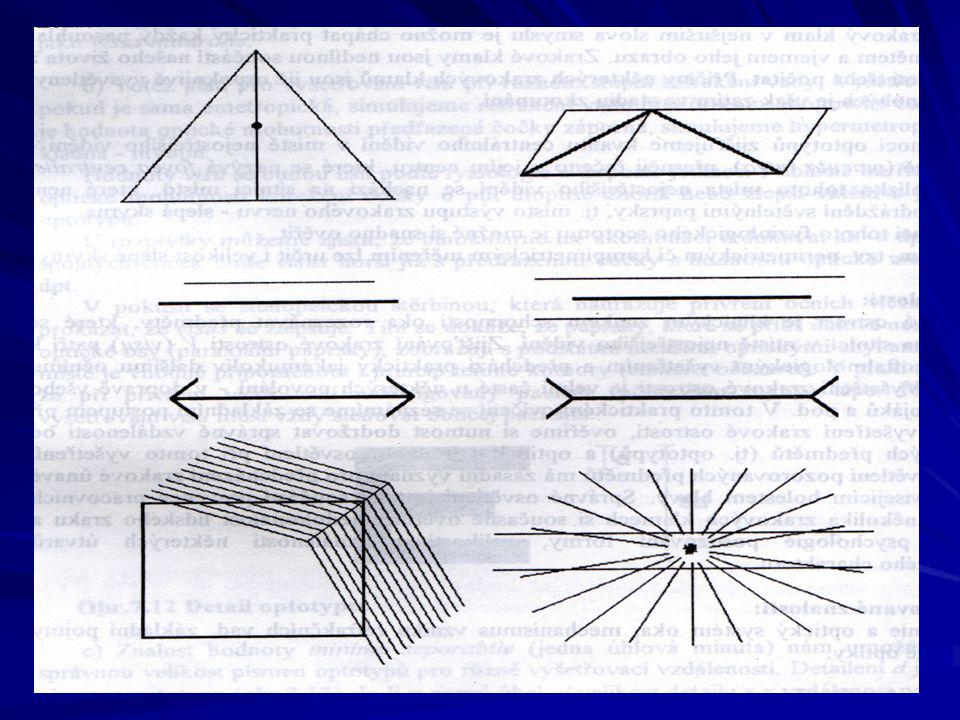
Zrakové klamy ukazují na úlohu zrakové části mozkové kůry při zpracování zrakové informace

35
Elektrické projevy sítnice
Elektrická aktivita sítnice je v úzkém vztahu k fotochemickým reakcím, probíhajícím ve fotoreceptorech při dopadu světla. raný receptorový potenciál pozdní receptorový potenciál Elektroretinografie (ERG), snímání pomocí dvou unipolárních svodů, mV
, snímání pomocí dvou unipolárních svodů, mV.")
36
Barevné vidění

37
Barevné vidění Barvy dělíme: základní
doplňkové, tj takové, které vzájemným smísením dají počitek neutrální šedé a bílé barvy. Každá vnímaná barva je charakterizována barevným tónem, světlostí a sytostí. barevný tón je určen vlnovou délkou světla, světlost intenzitou světla sytost barevností počitku. J. E. Purkyně změna poměrné světelnosti barev při adaptaci oka na tmu – PURKYŇŮV JEV

38
Barevný trojúhelník CIE
x – červená b. 650 nm, y – zelená b. 530 nm z – modrá b. 460 nm x + y + z = 1

39
Barevné vidění – spektrální citlivost
Čípky citlivé na červenou barvu Čípky citlivé na modrou barvu Čípky citlivé na zelenou barvu

40
Citlivost oka na vlnovou délku
Čípky „sumárně“

41
Barvocit Monochromáti - vnější svět vnímají pouze v odstínech šedi
Schopnost správného vnímání barev lidským okem Mechanismus vnímání barev není sice ještě jednoznačně rozřešen, všeobecně je však přijímána tzv. trichromatická teorie, spojená se jmény Helmholtze, Lomonosova a Younga . Jednotliví autoři se liší jen v charakteristice tří základních barev. Helmholtz za ně považoval červenou, zelenou a fialovou, Lomonosov a Young červenou, žlutou a modrou. Monochromáti - vnější svět vnímají pouze v odstínech šedi Dichromáti – částečná ztráta barvocitu, v sítnici chybí mechanismus pro vnímání jedné ze základních tří barev Trichromáti – jedinci s normálním barvocitem

42
Vyšetřování barvocitu
Pseudoizochromatické tabulky různých autorů (Stillingovy, Velhagenovy, Ischiharovy, Rabkinovy). Číslice nebo písmena jsou sestavena z okrouhlých barevných políček v záměnné barvě. Dichromát daného typu písmeno či číslici nerozezná. Vyšetření anomaloskopické. Nagelův anomaloskop je modifikovaný spektrální fotometr, pomocí něhož se barvocit určuje ze vztahu vyšetřovaného k vidění červené a zelené barvy.
. Číslice nebo písmena jsou sestavena z okrouhlých barevných políček v záměnné barvě. Dichromát daného typu písmeno či číslici nerozezná. Vyšetření anomaloskopické. Nagelův anomaloskop je modifikovaný spektrální fotometr, pomocí něhož se barvocit určuje ze vztahu vyšetřovaného k vidění červené a zelené barvy.")
43
Meze lidského zraku: zraková ostrost - testuje se pomocí
Snellenových optotypů (viz praktika) - dána úhlem jedné obloukové minuty limit citlivosti: 2-3 fotony během několika milisekund kritická frekvence splývání světelných impulsů: Hz v závislosti na jasu omezení vlnovými délkami světla: nm mez stereoskopického vidění: rozdíl stereoskopické paralaxy menší než dvacet úhlových vteřin
 - dána úhlem jedné obloukové minuty. limit citlivosti: 2-3 fotony během několika milisekund. kritická frekvence splývání světelných impulsů: Hz v závislosti na jasu. omezení vlnovými délkami světla: nm. mez stereoskopického vidění: rozdíl stereoskopické paralaxy menší než dvacet úhlových vteřin.")
44
Autoři Vojtěch Mornstein, Lenka Forýtková Obsahová spolupráce: Ivo Hrazdira, Carmel J. Caruana Grafika: - Poslední revize: Březen 2012

Podobné prezentace


 Mgr. Martin Šmíd.>")


